|
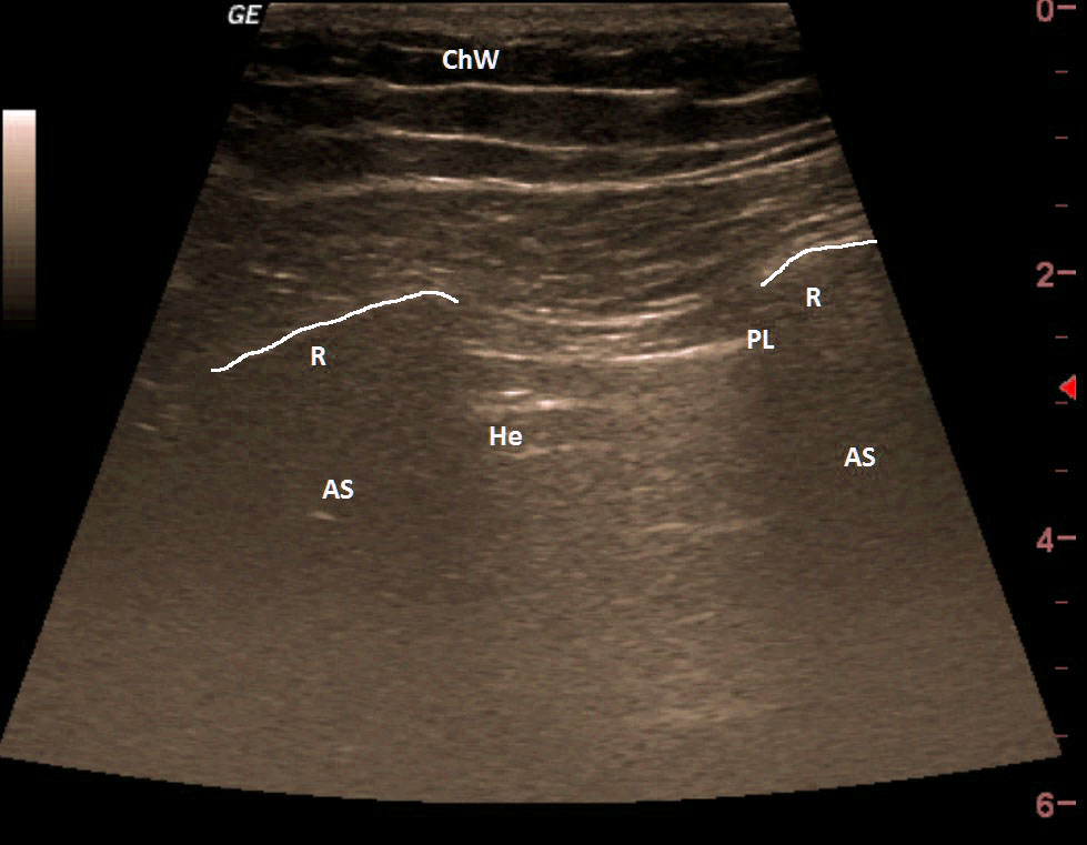
After placing the probe ventrally around the midclavicular line, we see the „bat sign“ (linear probe), which disappears during respiratory excursion and is replaced by the image of the heart. Be careful - this is not the „lung point“, which can be seen in a pneumothorax.
| ChW | - | Chest Wall |
| PL | - | Pleural Line |
| A | - | A line |
| He | - | Heart |
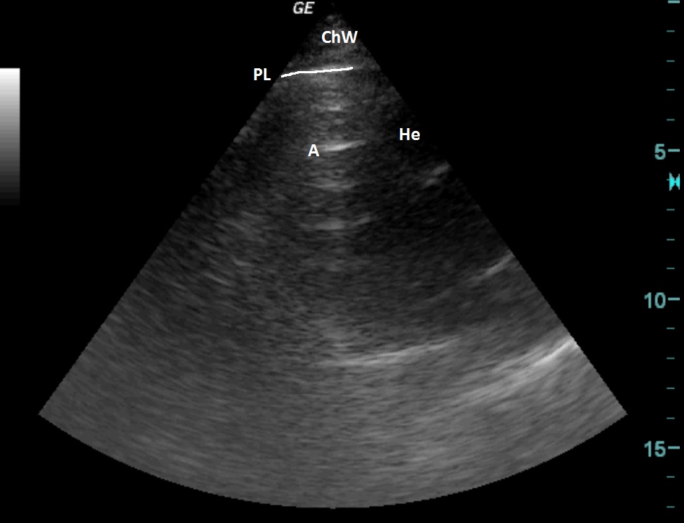
A similar picture also with a sector probe.
| ChW | - | Chest Wall |
| PL | - | Pleural Line |
| A | - | A line |
| He | - | Heart |
 česky
česky english