Sono Atlas

   |   

Your browser does not support images in SVG format, please update your browser.

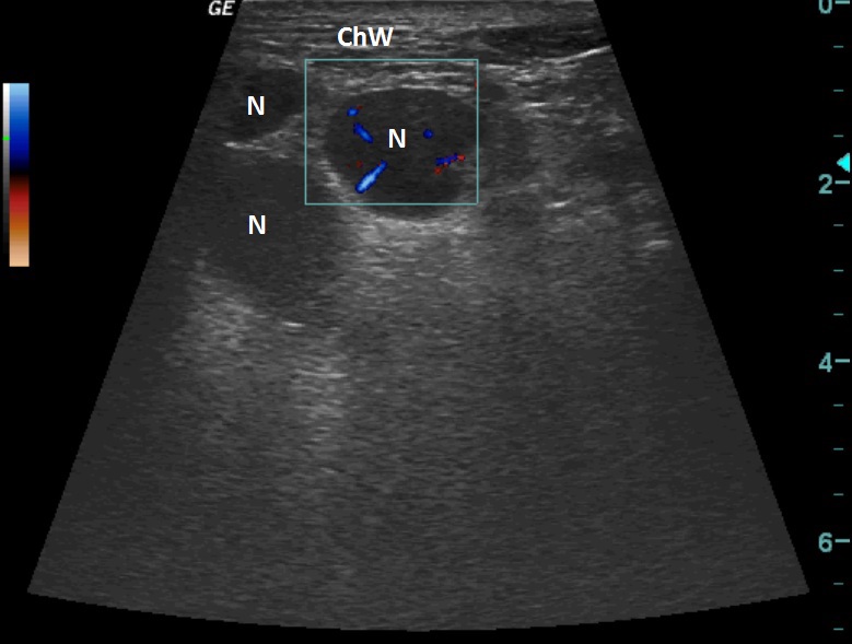
A patient with small cell lung cancer; in the left supraclavicular area a palpable mass.


On the ultrasound we can see three oval hypoechoic structures, for one of them Color Flow Doppler was used to view the vasculature. 

ChW - Chest Wall
N - Node


RTG
 

  © MUDr. Rastislav Šimek, 2013

  Project Partner:
Logo GlaxoSmithKline

X-ray and CT documentation used - Department of Imaging Methods, Tomas Bata Regional Hospital, JSC, Zlín.
All materials used on this site are for educational purposes and their use, modification, distribution or use in any other way either partially or completely without the prior written consent of the author is prohibited.