|
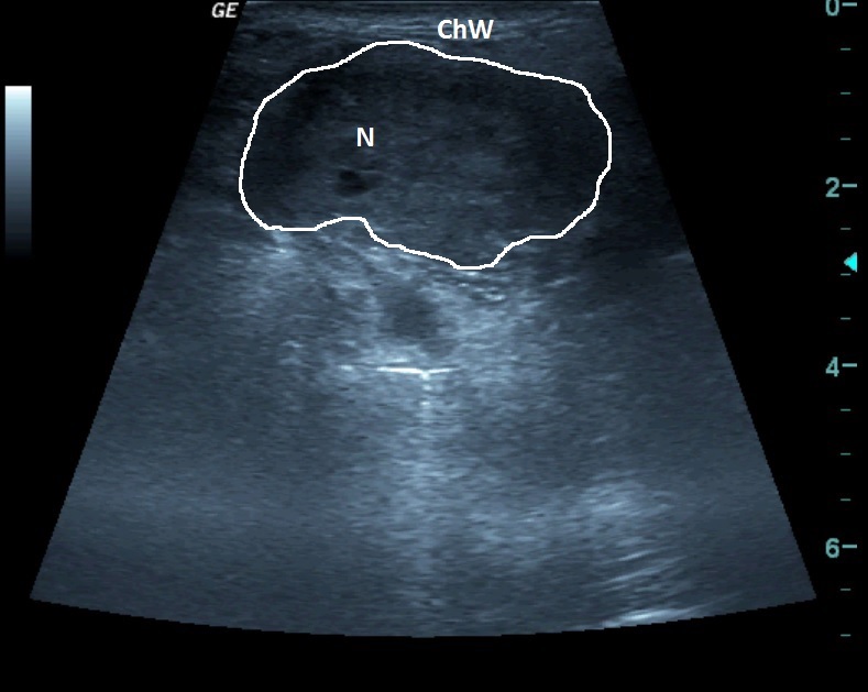
Patients with small cell cancer; in the left supraclavicular area a palpable mass.
At the ultrasound examination an irregular oval structure about 2x4 cm large – a node - can be seen.
| ChW | - | Chest Wall |
| N | - | Node |
Patients with small cell cancer; in the left supraclavicular area a palpable mass.
At the ultrasound examination an irregular oval structure about 2x4 cm large – a node - can be seen.
| ChW | - | Chest Wall |
| N | - | Node |
© MUDr. Rastislav Šimek, 2013
Project Partner:![]()
 česky
česky english