|
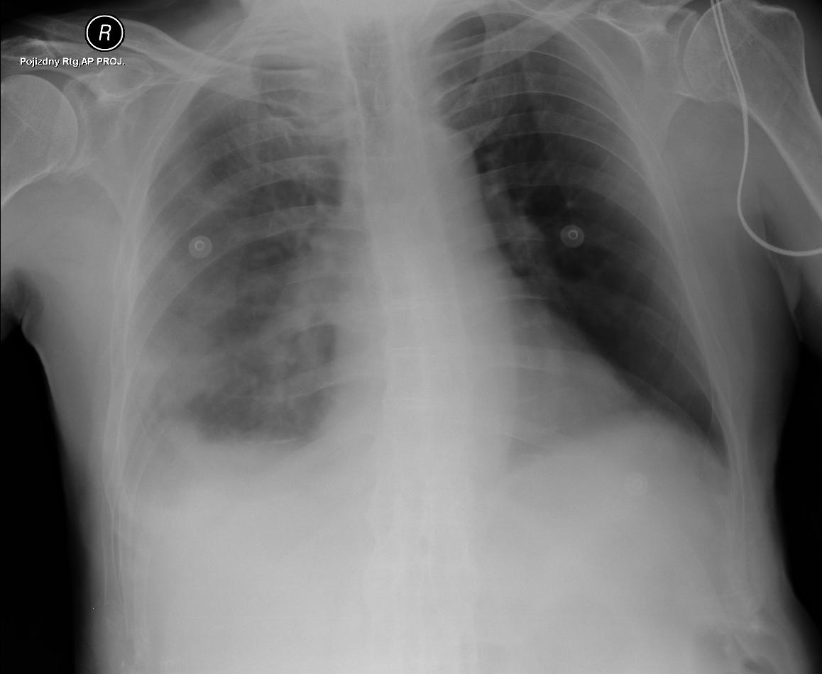
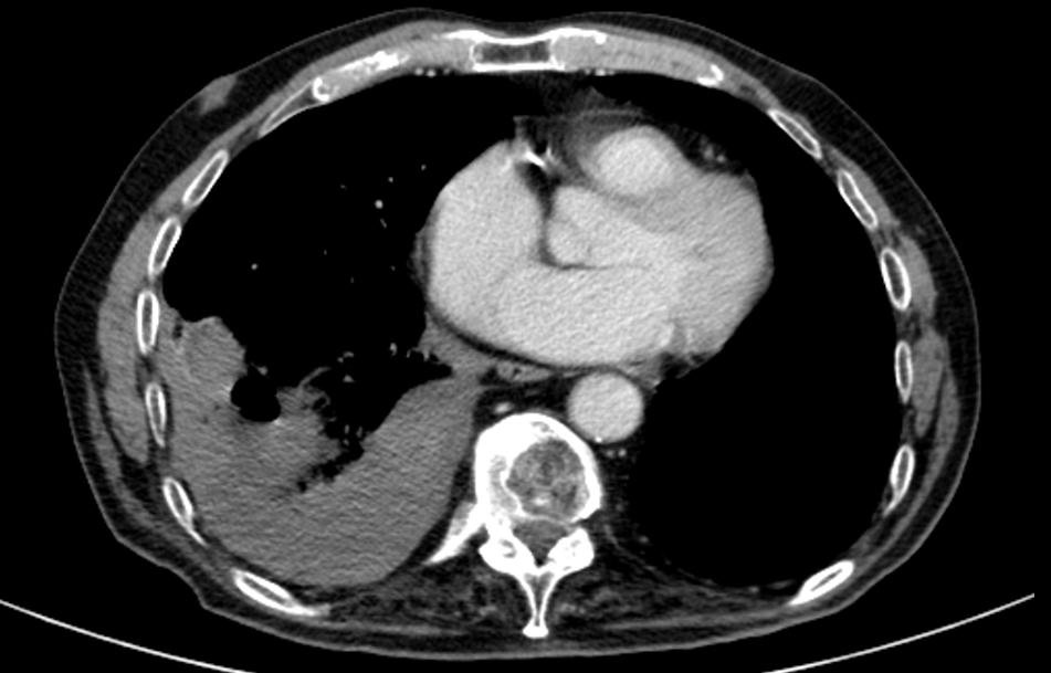
A patient with unclear non-regressing finding in the right lung, according to CT in the right lower lobe in S9 peripherally large irregular condensation of nodular contours in places, multiple spiculation around the area, isolated minor calcification. After a biopsy of the lodes hematoma developed.
At the ultrasonographic examination immediately after the biopsy wavy hyperechoic structures press down on the diaphragm - hematoma.
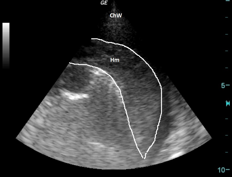
| ChW | - | Chest Wall |
| Hm | - | Hematoma |
 česky
česky english