|
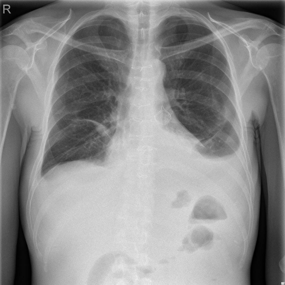
A patient after having had heart surgery for pulmonary and tricuspid valve disease; treated with bioprosthesis. After the operation, development of postpericardiotomy syndrome - recurrent fluidothorax on the left.
The ultrasound examination showed hyperechoic fluidothorax on the left with compressive atelectasis in part of the lung. A pleural puncture resulted in straw yellow effusion, biochemical of transudate character, cytology mesothelial effusion.
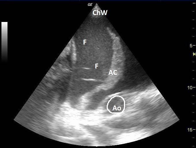
| ChW | - | Chest Wall |
| F | - | Fluidothorax |
| AC | - | Compressive Atelectasis of the Lung |
| Ao | - | Aorta |
 česky
česky english