|
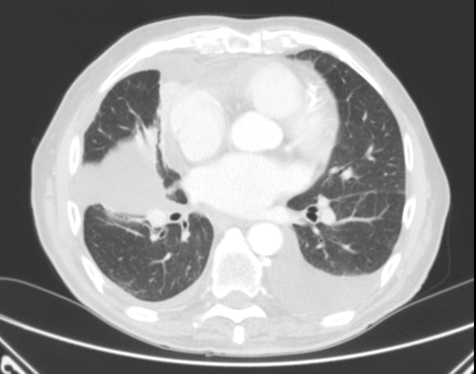
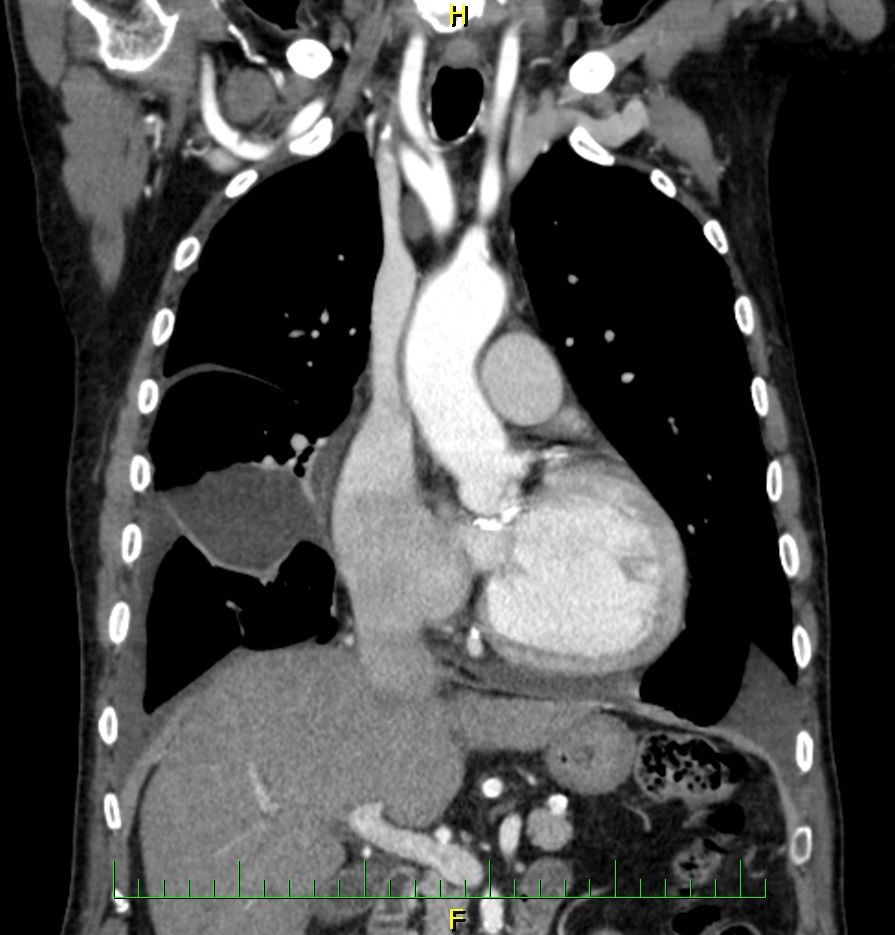
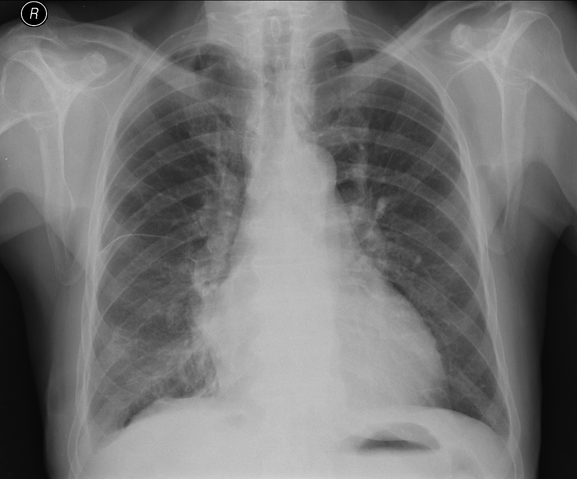
A patient with bilateral effusion, on the right it is interlobar.
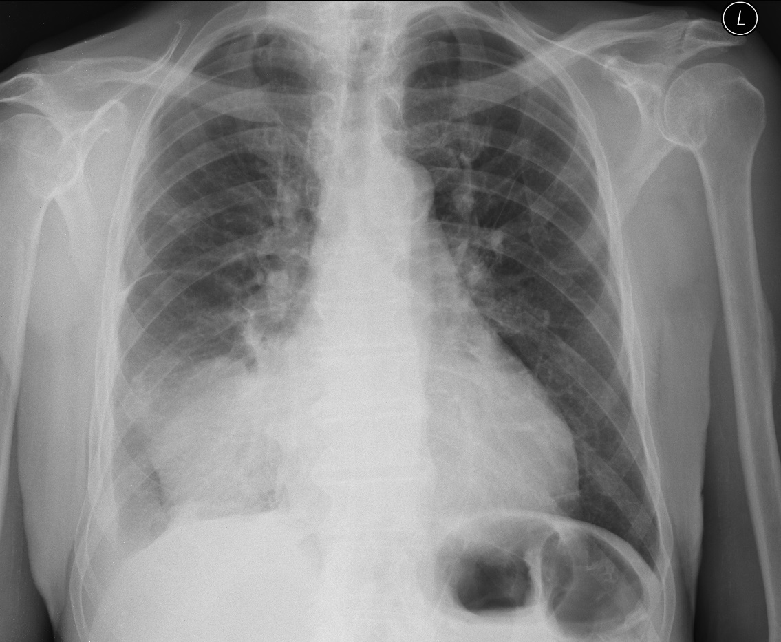
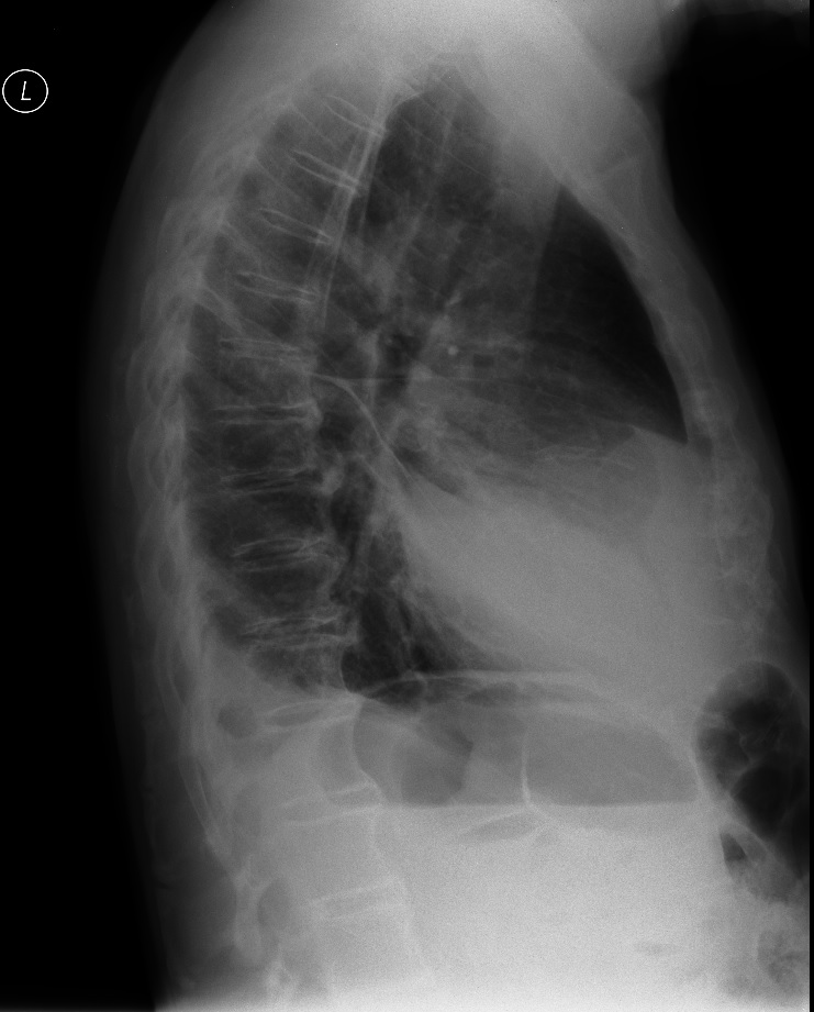
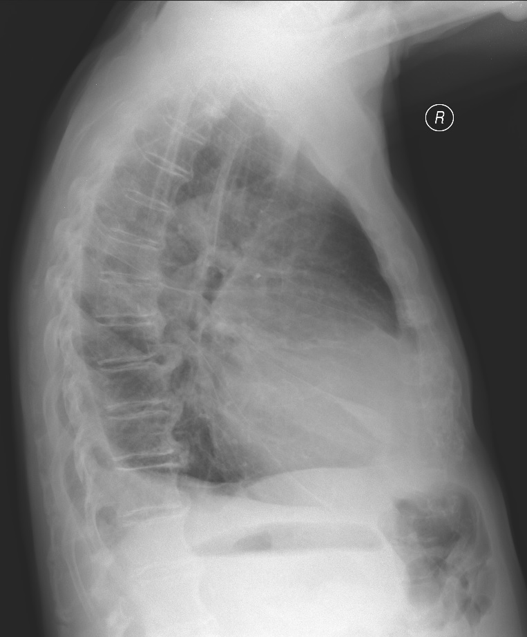
On the ultrasonographic examination via the lateroventral approach an encapsulated hypoechoic collection can be seen on the right; a pleural puncture of this place was carried out and 200 ml straw yellow effusion collected. Cytology showed an inflammatory modified exudate without the presence of malignant cells. The check-up chest X-ray showed a virtually complete regression of the interlobar effusion.
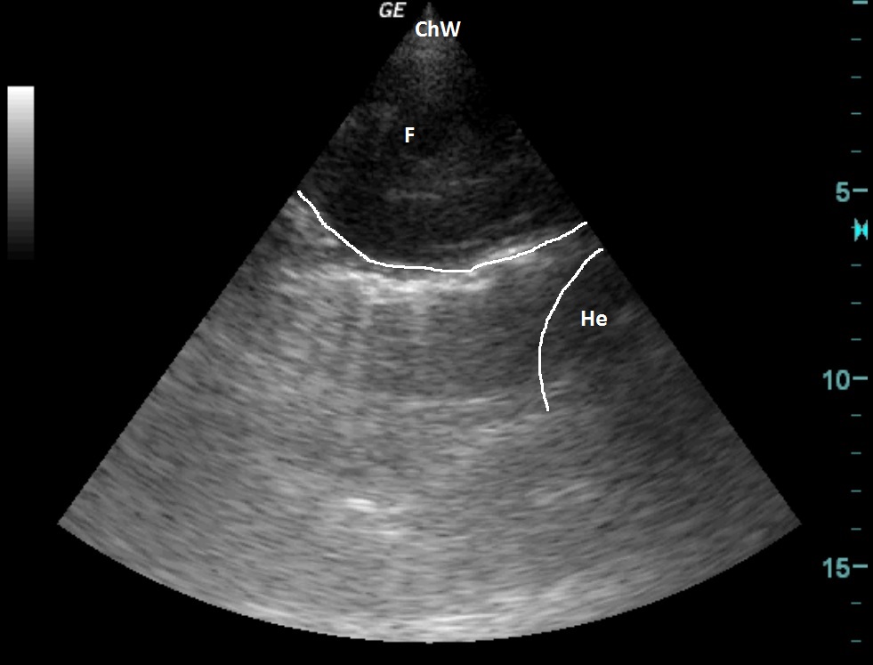
| ChW | - | Chest Wall |
| F | - | Fluidothorax |
| He | - | Heart |
After the puncture
 česky
česky english