|
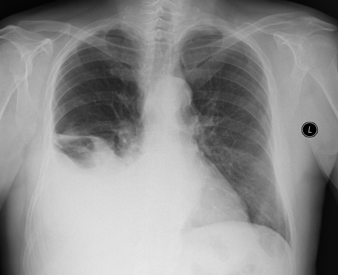
A patient with a central tumour in the hilum of the right lung with atelectasis, fluidothorax on the right, pleural metastases, hilar and mediastinal lymphadenopathy, bilateral metastasis in the left adrenal gland.
The ultrasound examination shows a relatively large fluidothorax, in the basal liver there are irregular oval hyperechoic structures - metastases.
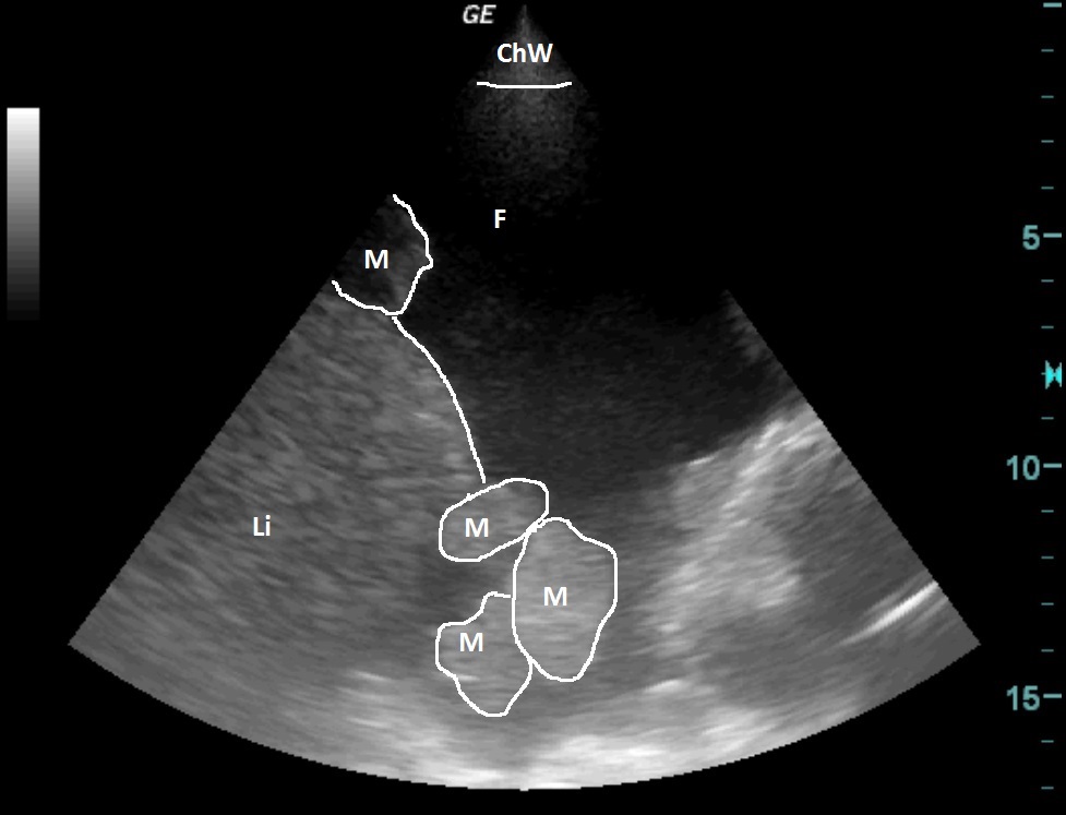
| ChW | - | Chest Wall |
| F | - | Fluidothorax |
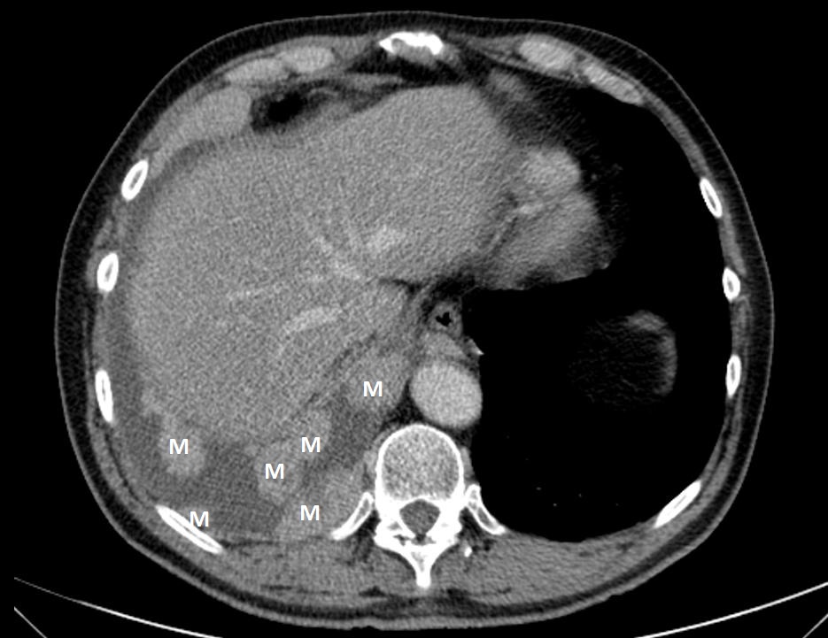
| M | - | Metastases |
| Li | - | Liver |
 česky
česky english