Sono Atlas

   |   

Your browser does not support images in SVG format, please update your browser.

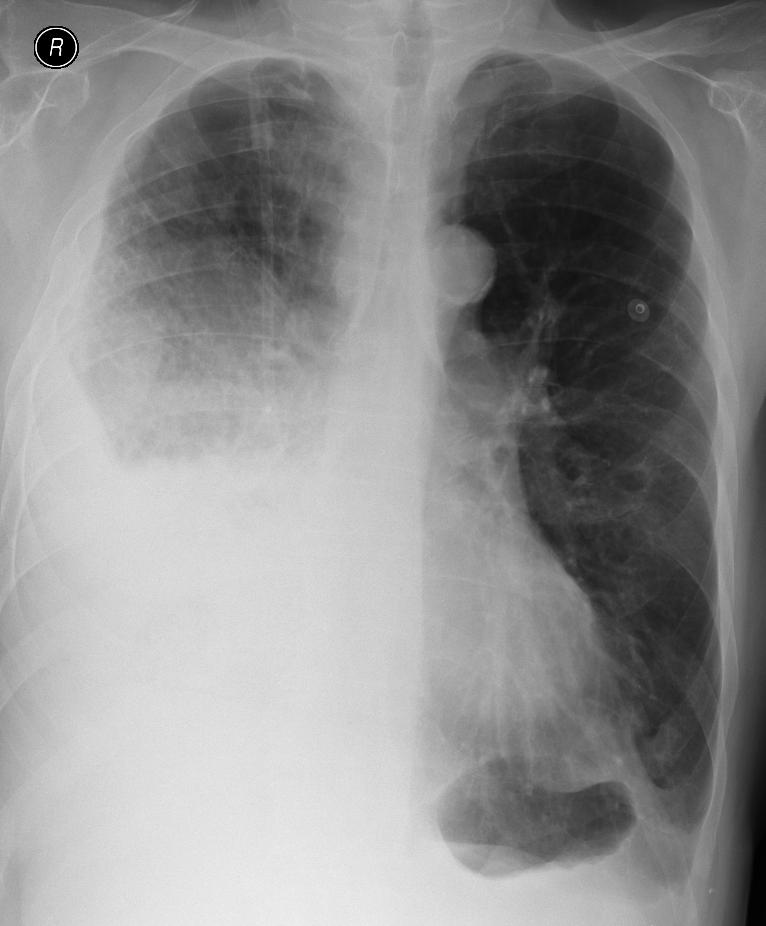
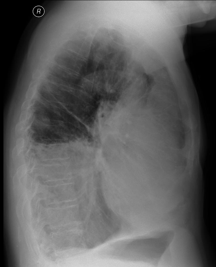
A patient with  right-side pneumonia, fluidothorax on the right.


On the ultrasound examination, we see fluidothorax, followed by alveolar consolidation with a noticeable dynamic air bronchogram (white arrow); the air in the bronchi acts as a contrast and moves in their lumen when the patient is breathing.
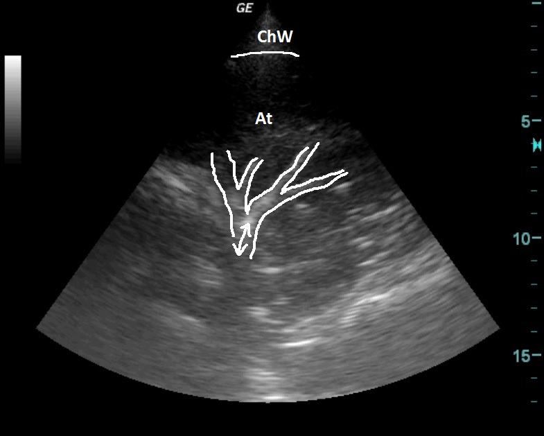


RTG
RTG

SONO

ChW - Chest Wall
At - atelectasis with a noticeable air bronchogram


  © MUDr. Rastislav Šimek, 2013

  Project Partner:
Logo GlaxoSmithKline

X-ray and CT documentation used - Department of Imaging Methods, Tomas Bata Regional Hospital, JSC, Zlín.
All materials used on this site are for educational purposes and their use, modification, distribution or use in any other way either partially or completely without the prior written consent of the author is prohibited.