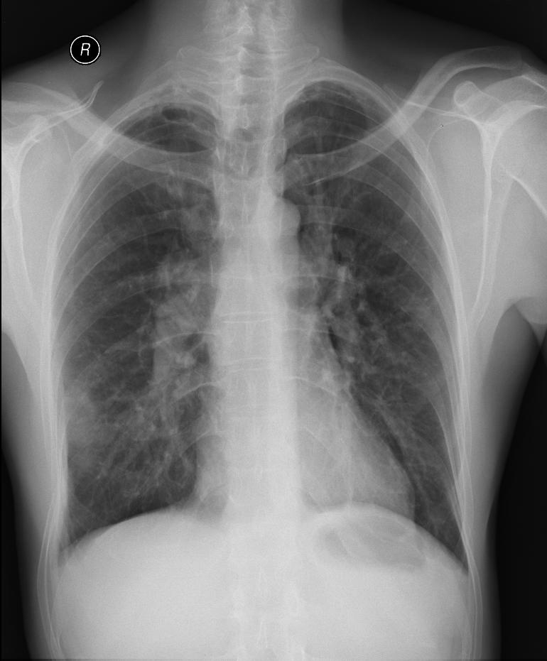
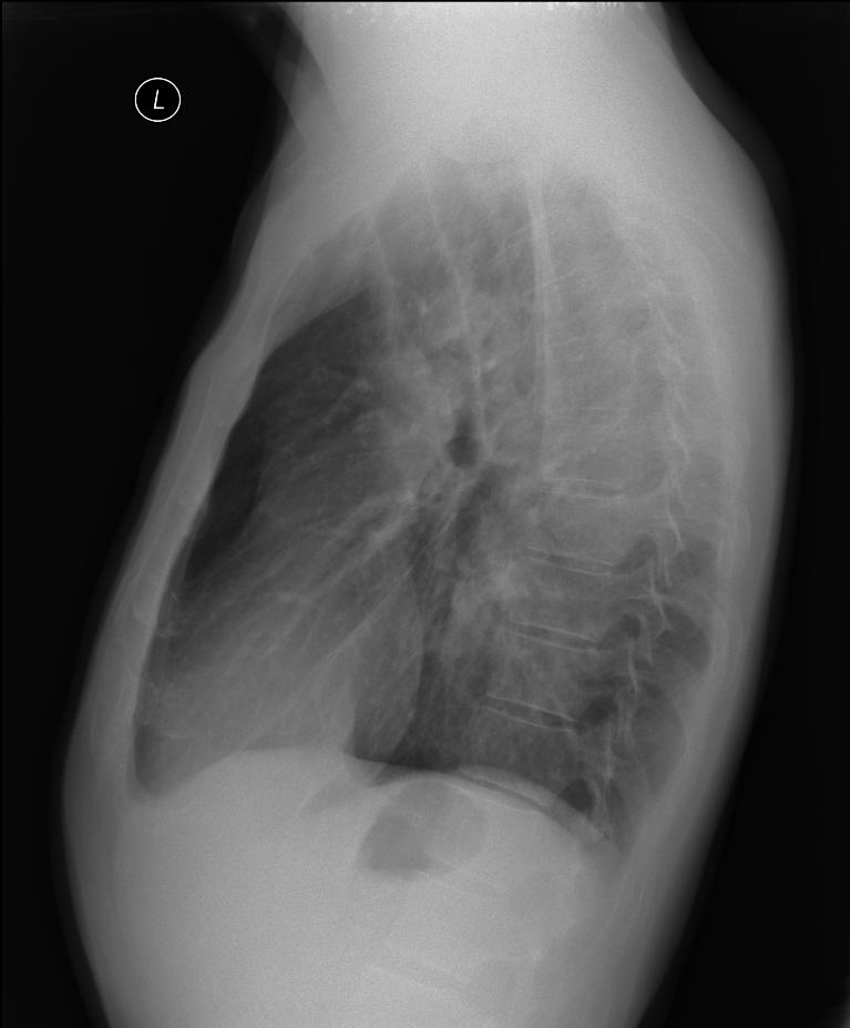
A patient with a history of pulmonary embolism on the left in the past, undergoing anticoagulant therapy. He was sent from a pulmonary clinic due to high temperatures, hemoptysis, and pain in the right hemithorax. On chest X-ray suspected infiltration on the border of the right middle and lower lung fields.
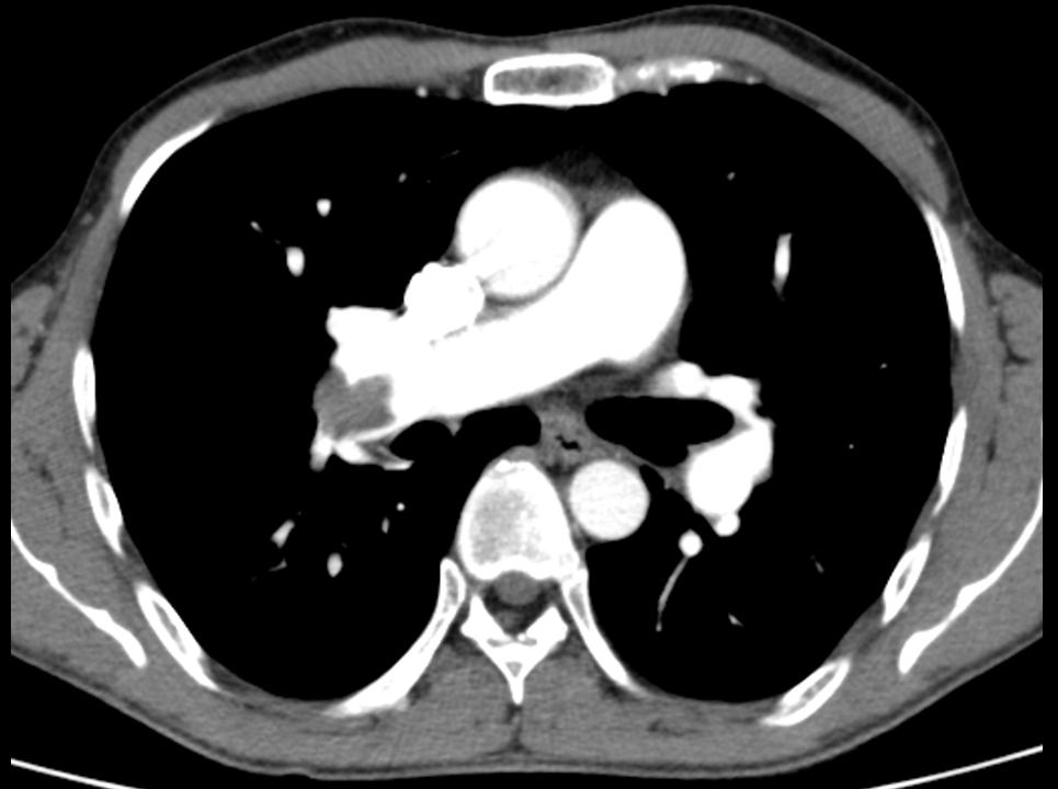
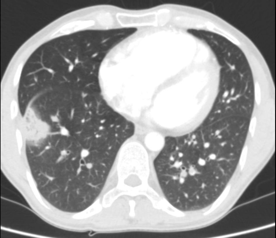
Ultrasound examination performed for the purpose of differential diagnosis, we see the disappearance of „lung sliding“, signs of alveolar consolidation – the „shred sign“, which happens in pneumonia. In this case colour Doppler was used, which showed alveolar consolidation in the area and no signs of vascular flow. Suspected pulmonary infarction (even given the medical history and clinical picture). The patient is sent for an acute CT angiogram, which confirms pulmonary embolism of the right main pulmonary artery branches, condensation in the periphery of the right, strongly suspected pulmonary infarction.
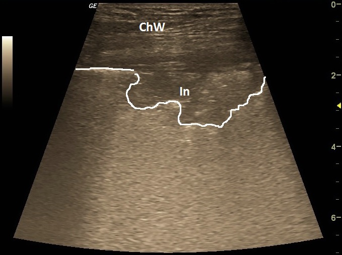
| ChW | - | Chest Wall |
| In | - | suspected pulmonary infarction |
 česky
česky english