|
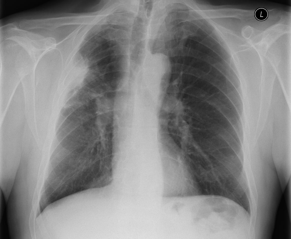
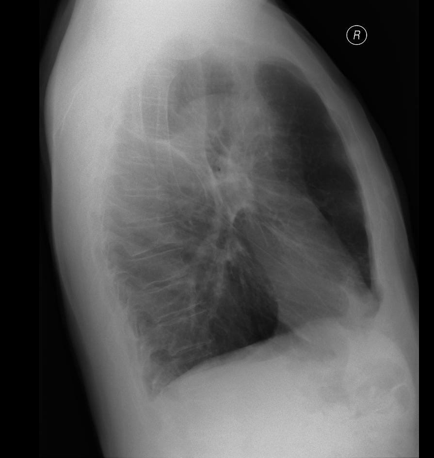
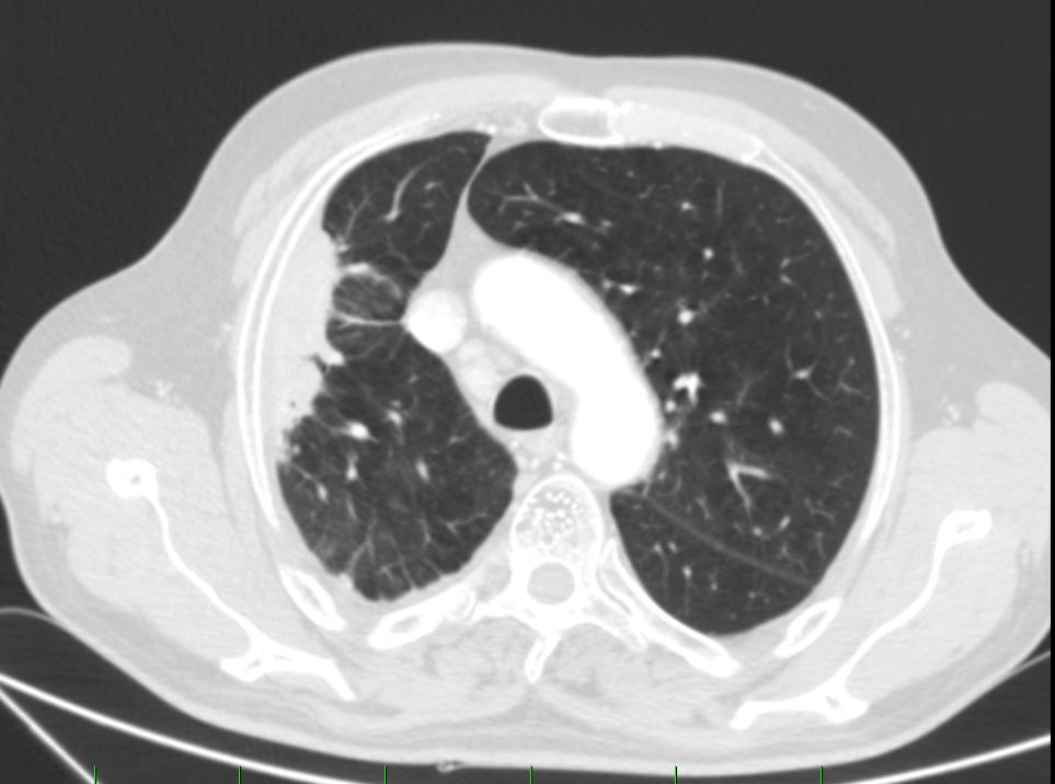
The patient was sent for further investigation due to non-regressive shaded area in the right middle and upper lung field according to RTG.
The ultrasonographic examination shows an irregular hypoechoic mass pressing down on the pleura, under ultrasound guidance anaesthesia performed, and we can see the anaesthetic leaking around the needle. Furthermore, a biopsy was performed with a true-cut needle in real time under ultrasound guidance. Histological examination confirms organizing pneumonia.
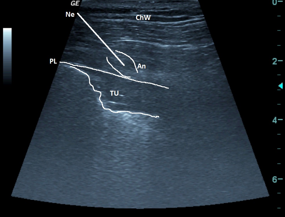
| ChW | - | Chest Wall |
| Ne | - | Needle |
| An | - | Anaesthetic |
| PL | - | Pleural Line |
| TU | Tumour |
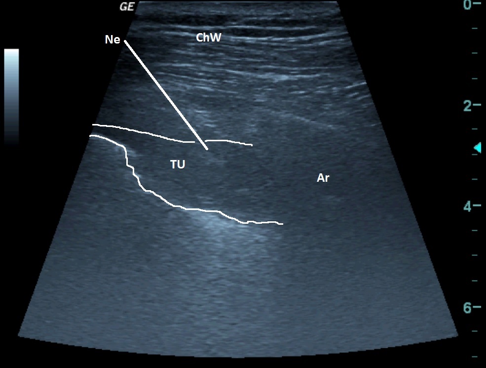
| ChW | - | Chest Wall |
| TU | - | Tumour |
| Ne | - | Needle |
| Ar | - | Artefact |
 česky
česky english