|
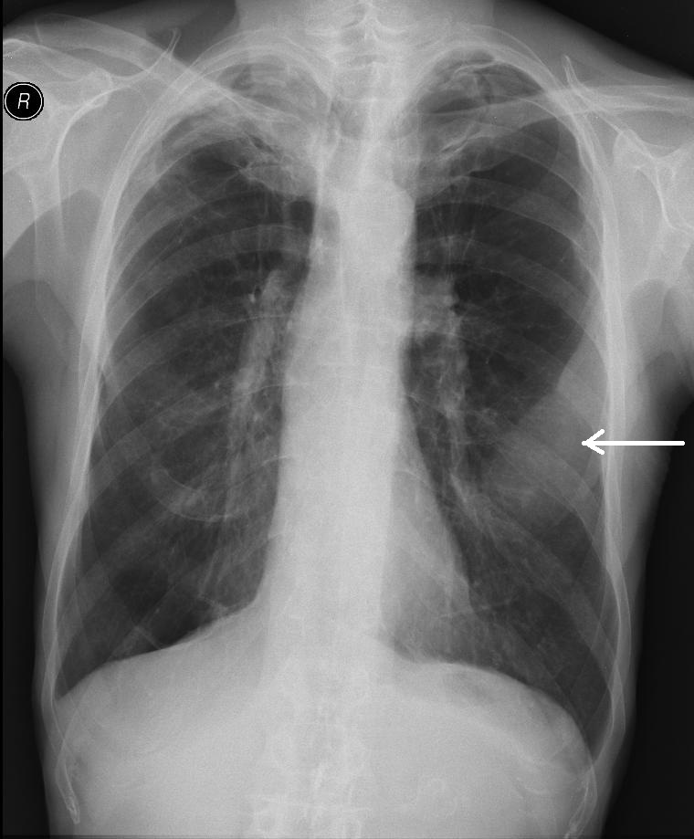
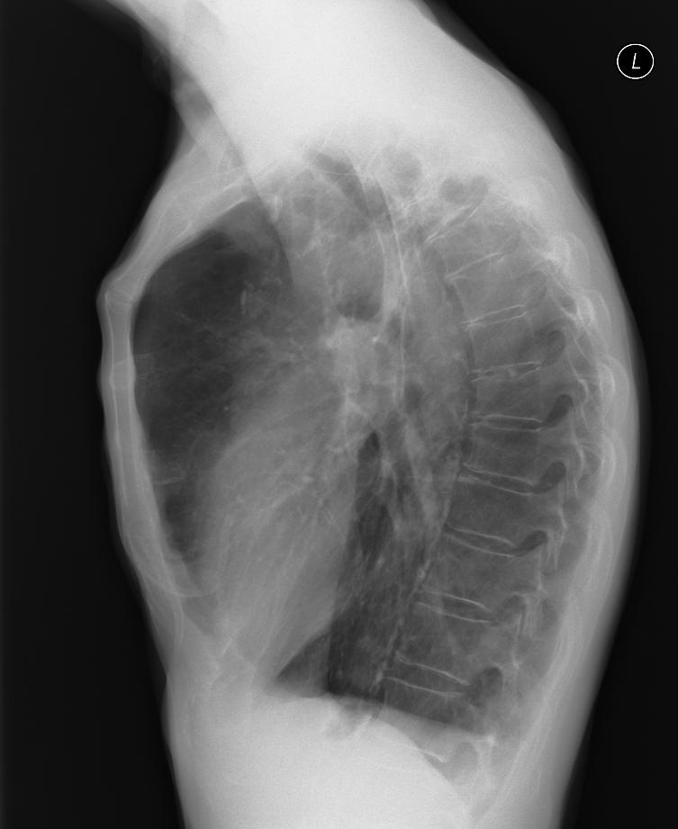
The patient is admitted to hospital due to pain in the left hemithorax, loss of appetite, weight loss. The X-ray shows a vague shadow on the left (arrow), infiltration considered.
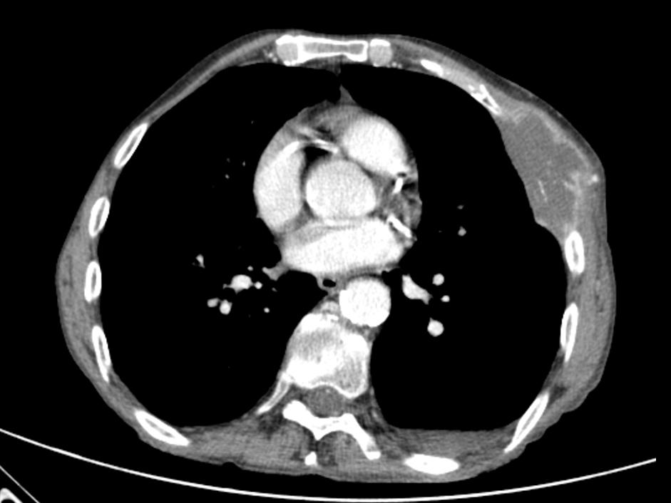
The ultrasound examination showed the presence of tumour masses in the chest wall, a biopsy with a true-cut needle was performed in real time; histological examination showed adenocarcinoma.
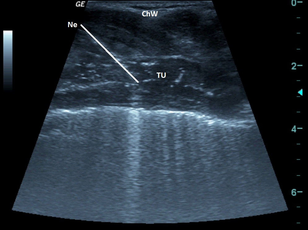
| ChW | - | Chest Wall |
| Ne | - | Biopsy Needle |
| TU | - | Tumour |
 česky
česky english