|
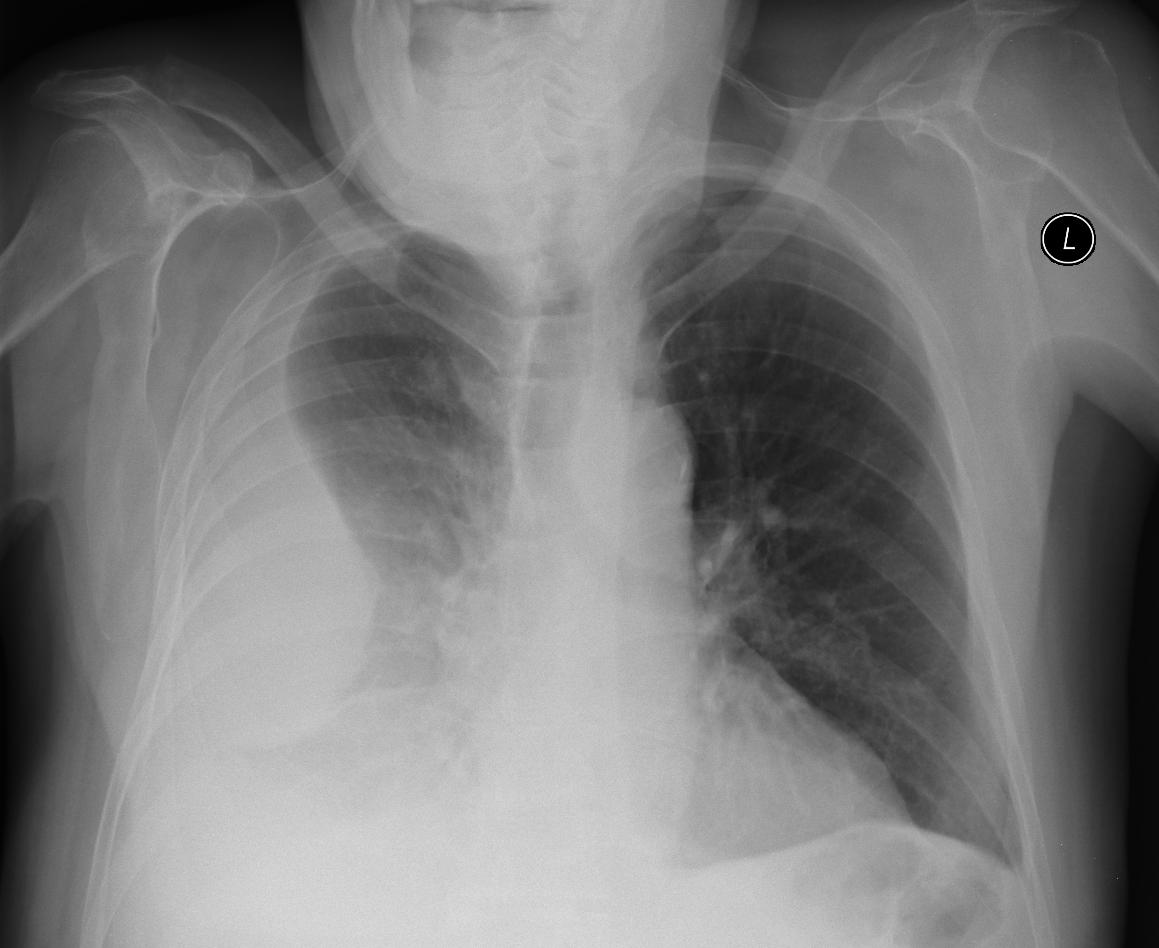
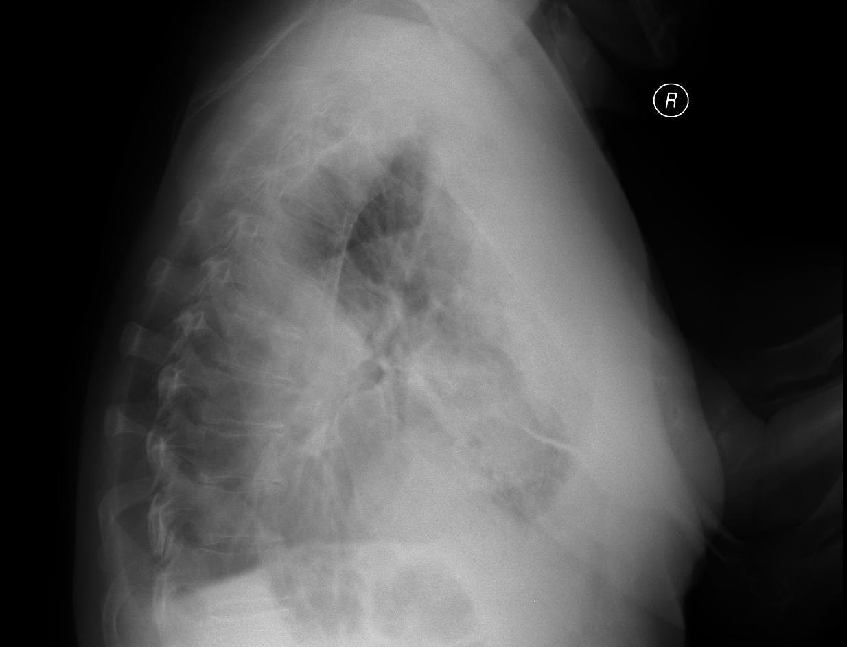
A patient with empyema on the right, chest drain inserted.
On the ultrasonographic examination residual fluid is shown, in which the drain can be seen. To be sure we can lightly pull on the drain from the outside, and the movement of the drain is seen in the pleural cavity. Suitable for confirmation of correct implementation when in doubt, or to avoid dislocation into the subcutaneous tissue or dislocation of the drain from the place where it should be.
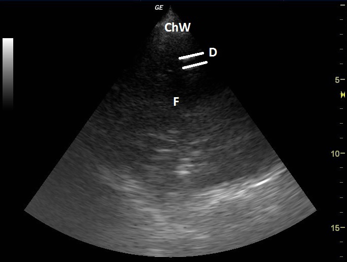
| ChW | - | Chest Wall |
| D | - | Drain |
| F | - | Fluidothorax |
 česky
česky english