|
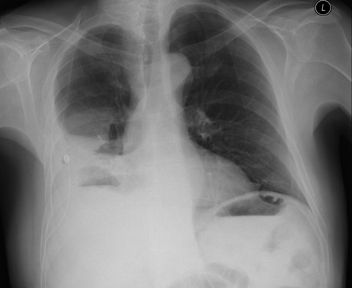
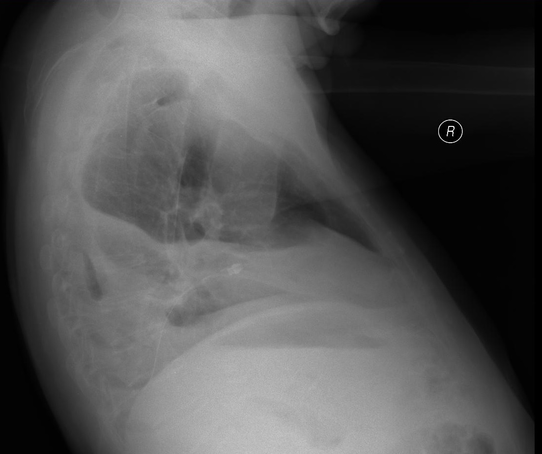
A patient with right thoracic empyema, sepsis, chest drain inserted under ultrasound guidance, purulent exudate removed, cytology confirmed empyema. In the following days the drain was flushed.
The ultrasonographic examination shows residual fluid, the flush was performed under ultrasound guidance in order to check the drain and check the effectiveness of the therapy. When the drain is being flushed we can see fluid leakage into the rest of the empyema cavity and how it fills up without any difficulties. Next the flushed fluid was suctioned out. The drain was not dislocated or blocked, and there was no fluid leakage into the subcutaneous tissue.
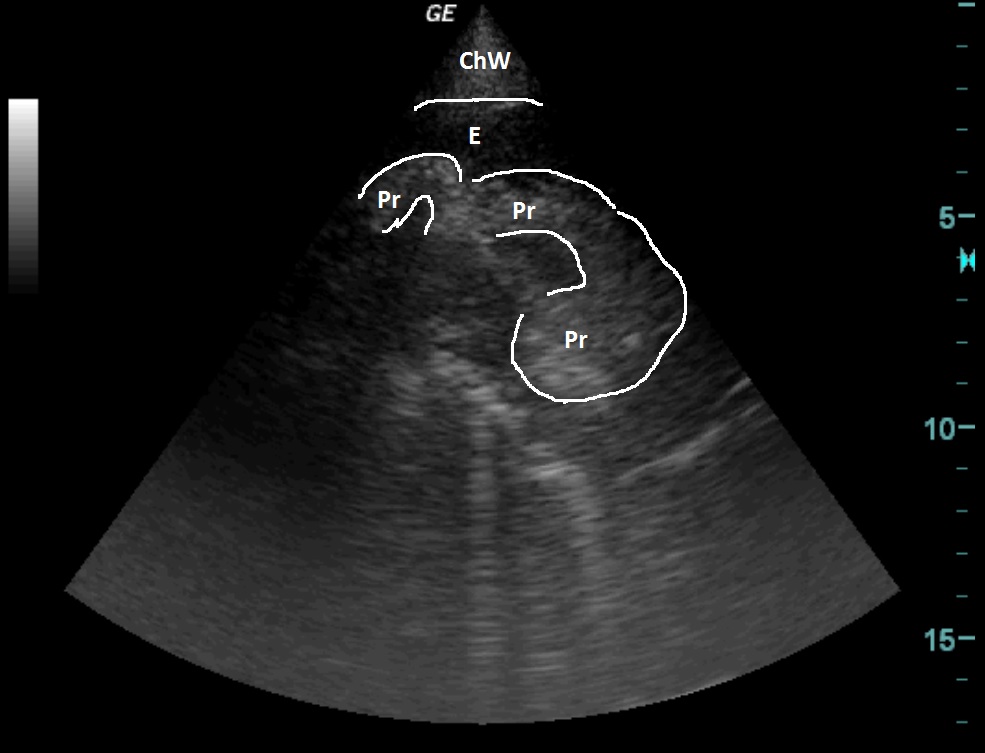
| ChW | - | Chest Wall |
| E | - | Empyema |
| PR | - | Flushing |
 česky
česky english