|
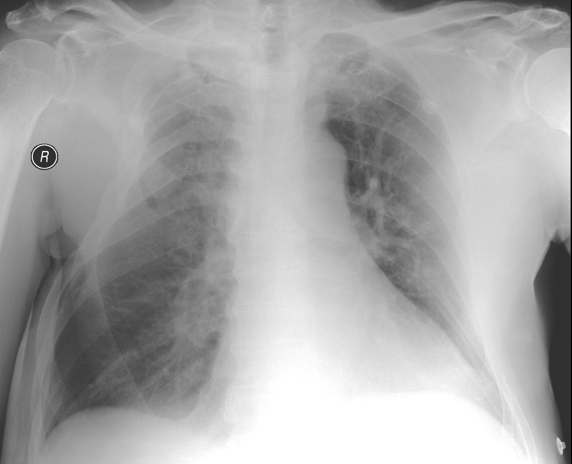
A 72-year-old patient who had purulent pleurisy on the right in childhood; since then there has been deformity of the chest. The Valsalva manoeuvre leads to lung herniation in the chest wall, and when coughing the herniation is accentuated even more.
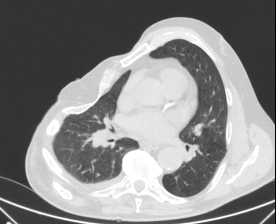
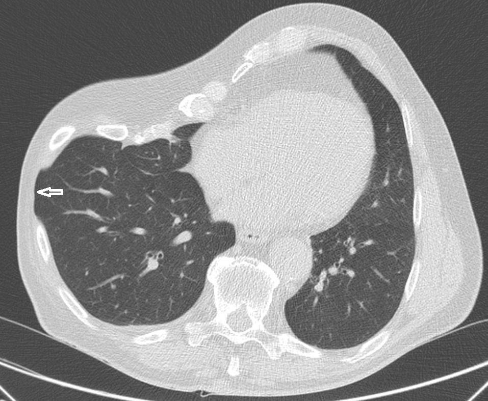
When the lungs are at rest we can see the „bat sign“, the pleural line, and „lung sliding“. When the patient is prompted to cough we can see the change in position of the pleural line towards the surface of the chest wall (herniation). During the Valsalva manoeuvre herniation was not noticeable. A similar picture, taken while at rest and during the Valsalva manoeuvre (arrow), was seen on CT.
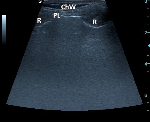
| ChW | - | Chest Wall |
| PL | - | Pleural Line |
| R | - | Rib |
 česky
česky english