|
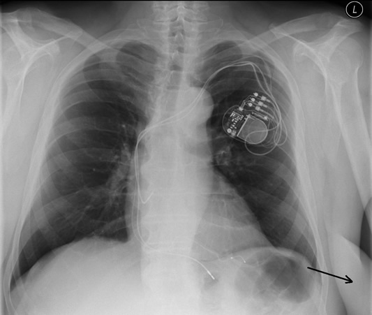
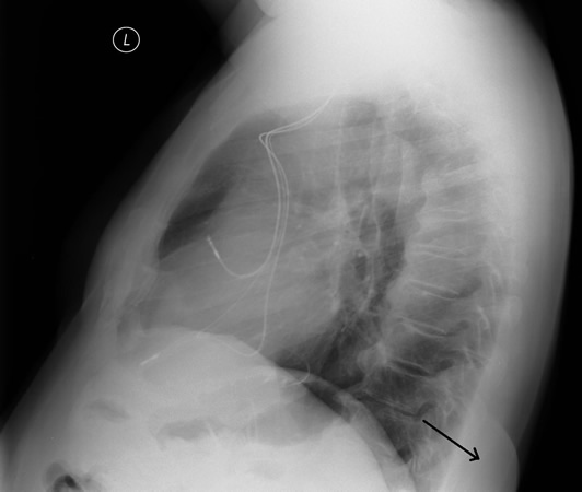
The patient was examined in the pulmonary clinic during the pre-operative examination – a large lipoma on the left side of the chest was removed.
An ultrasound examination was performed with a linear and a sector probe which were placed on the left in a dorsobasal position. A relatively homogeneous hyperechoic structure with hyperechoic septa can be seen. Regular adipose tissue with fibrous septa confirmed at histological examination.
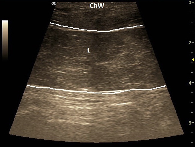
| ChW | - | Chest Wall |
| L | - | Lipoma |
| Lien | - | Spleen |
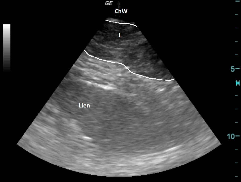
| ChW | - | Chest Wall |
| L | - | Lipoma |
| Lien | - | Spleen |
 česky
česky english