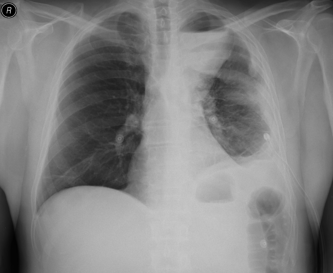
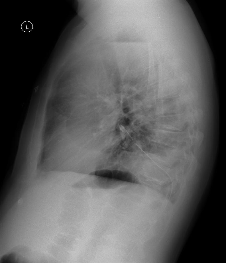
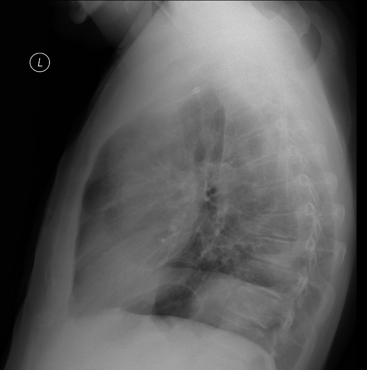
A patient with empyema on the left. Thoracic drainage on the left performed on the basal side, purulent exudate removed with the character of empyema. After drainage an encapsulated collection of surface fluid remains in the upper lung field.
The probe was attached to the left subclavian area; the ultrasound shows the encapsulated fluid collection, pressing down on the aortic arch. Under ultrasound guidance a puncture was performed to suction out the cloudy white-green fluid, after which a chest tube was inserted under the guidance of an ultrasound examination (complicated approach, aortic arch). Cytology confirmed empyema. After drainage there was a complete regression of the encapsulated collection.
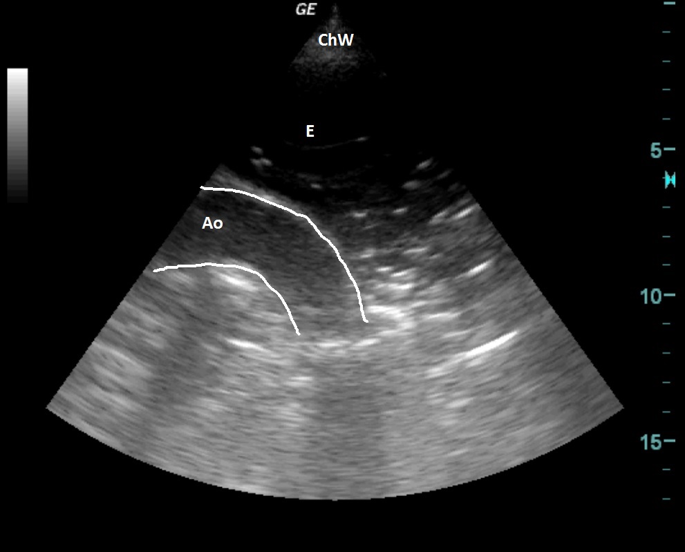
| ChW | - | Chest Wall |
| E | - | Empyema |
| Ao | - | Aorta - Aortic arch |
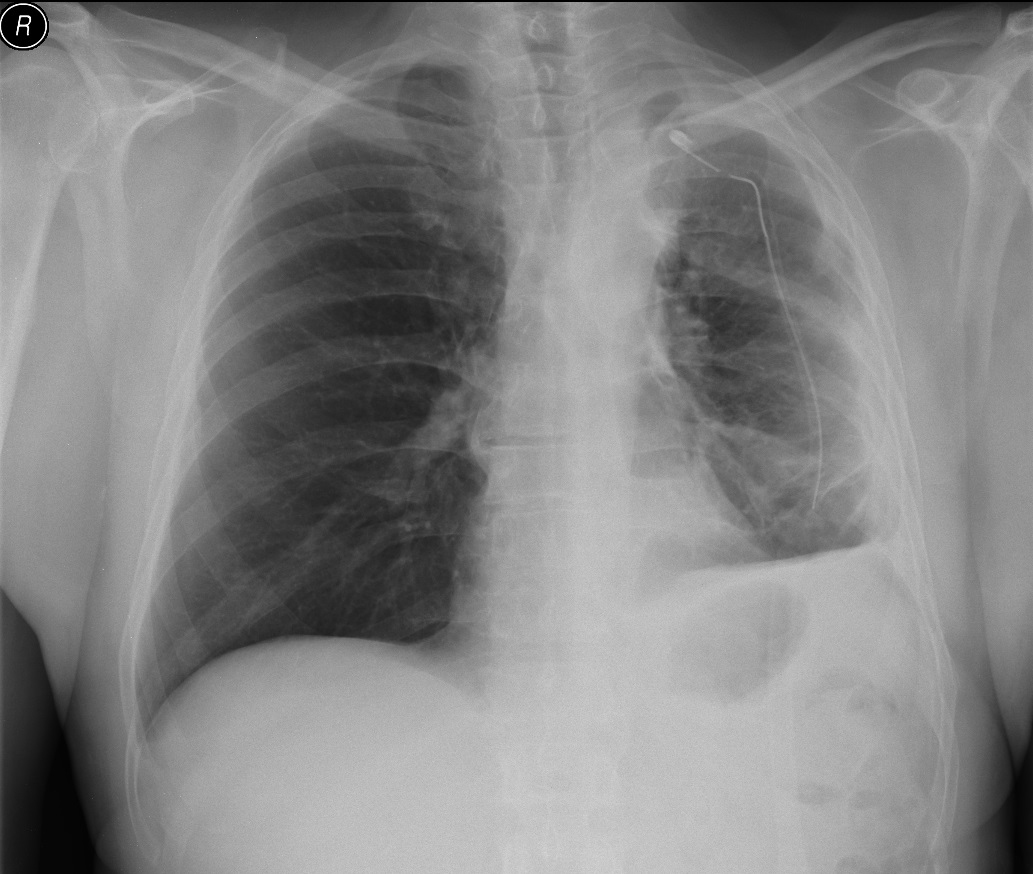
After drainage of the encapsulated upper fluid
 česky
česky english