|
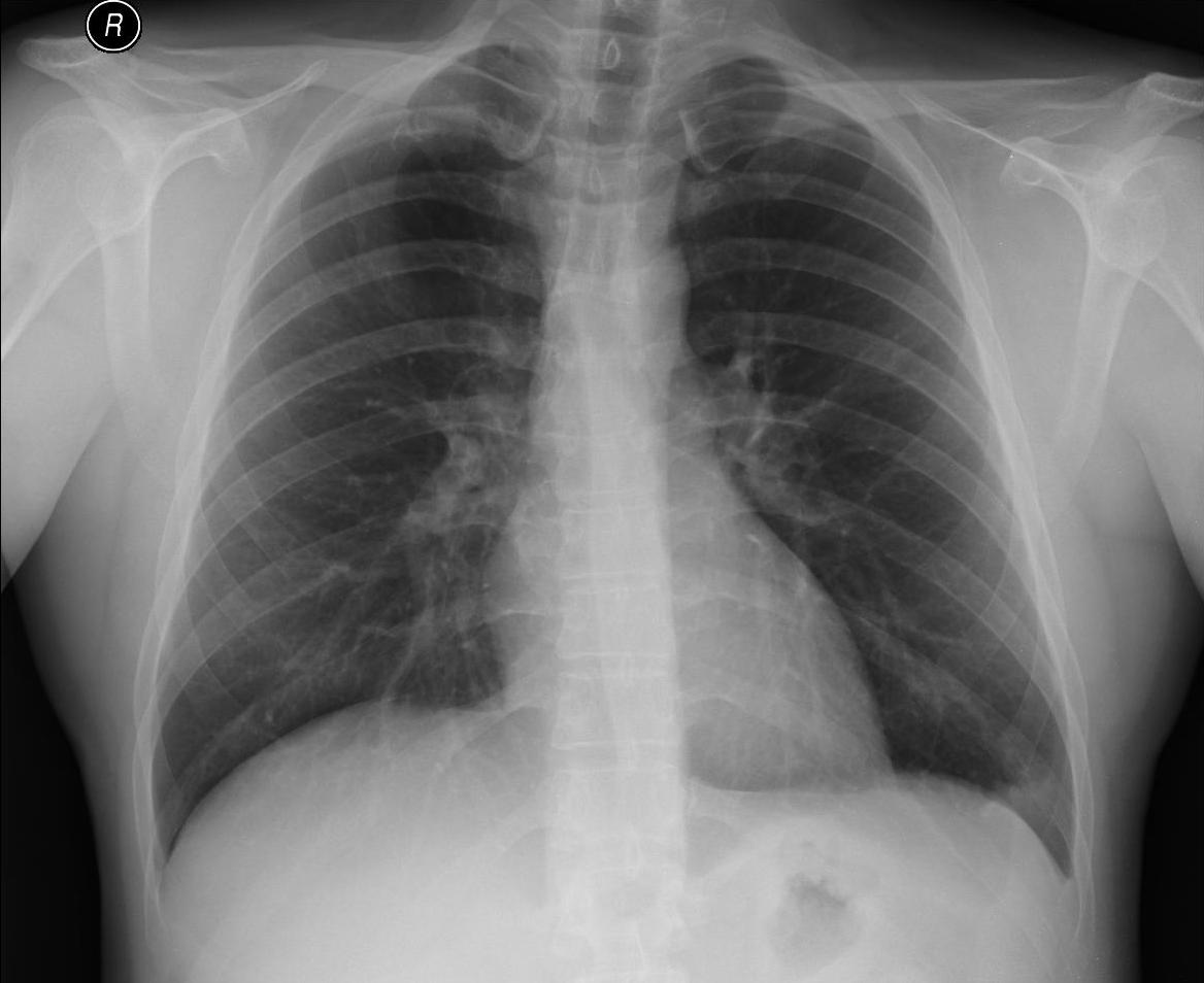
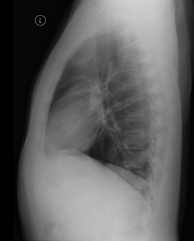
A patient with minor pneumonia in the left lower lobe (according to the chest X-ray small infiltration on the left outer diaphragmatic angle).
On the ultrasound examination a small effusion was shown, during respiratory excursions we can see the movement of pulmonary parenchyma in the area of the costodiaphragmatic angle, where there is even small fluidothorax (max. width of about 5 mm). No intervention.
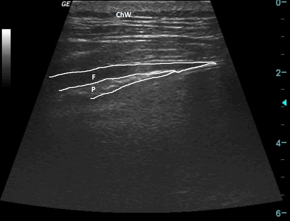
| ChW | - | Chest Wall |
| F | - | Fluidothorax |
| P | - | Pulmonary Parenchyma |
 česky
česky english