|
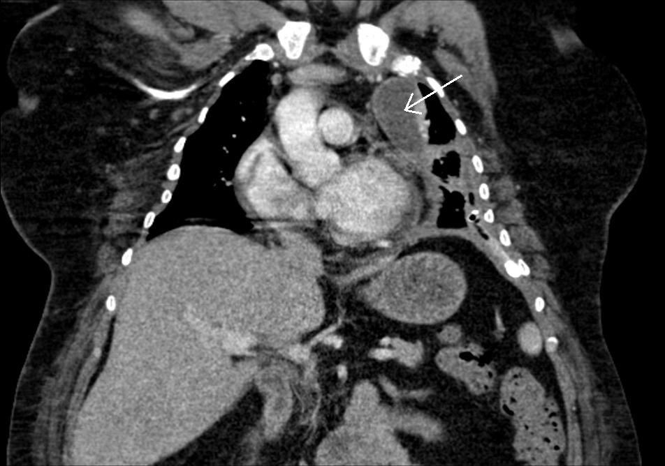
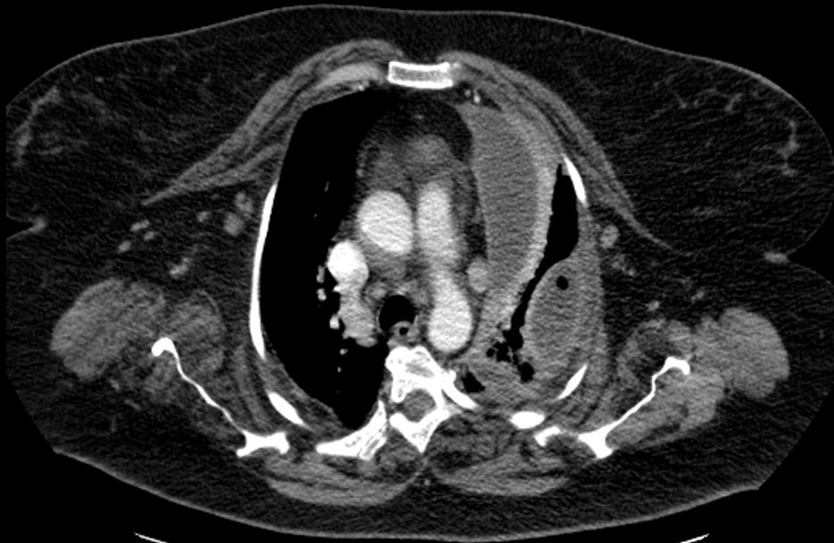
A patient with thoracic empyema on the left; after thoracic drainage encapsulated paracardial fluid collection persists.
The ultrasound examination carried out via the subclavian approach on the left shows the encapsulated fluid collection with septa pressing down on the heart. In the pericardium there is a small pericardial effusion.
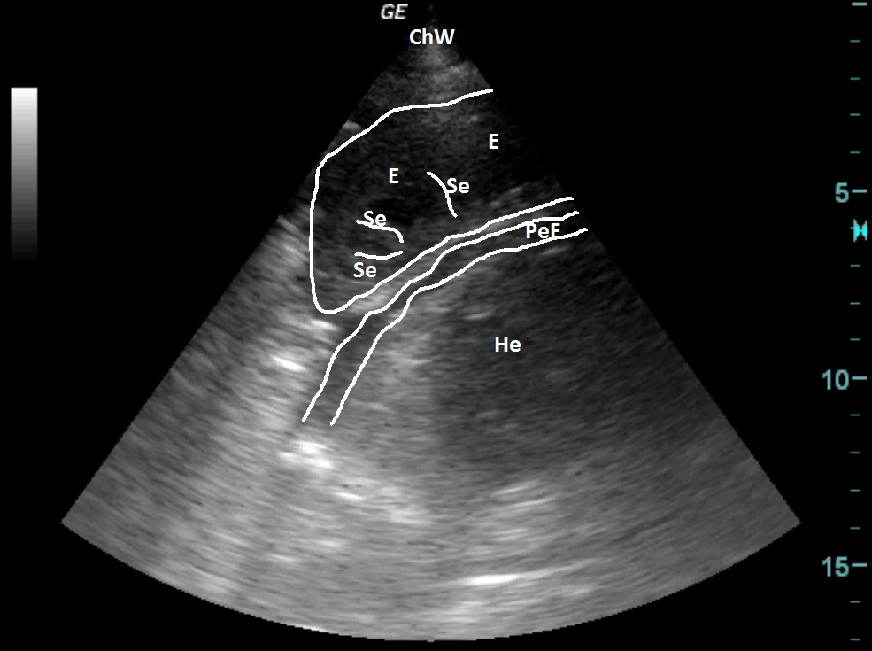
| ChW | - | Chest Wall |
| E | - | Empyema |
| Se | - | Septa |
| PeF | - | Pericardial Effusion |
| He | - | Heart |
 česky
česky english