|
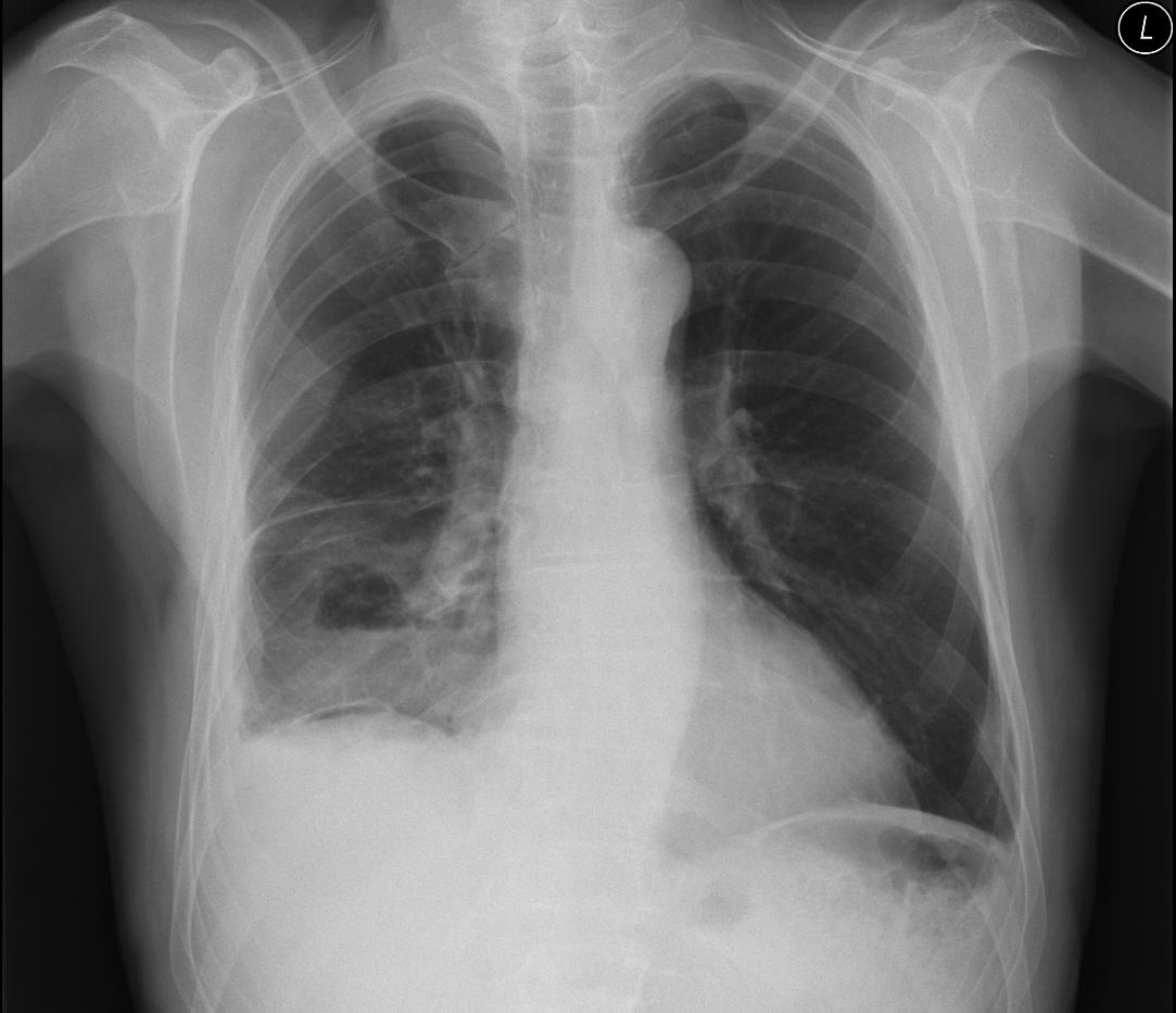
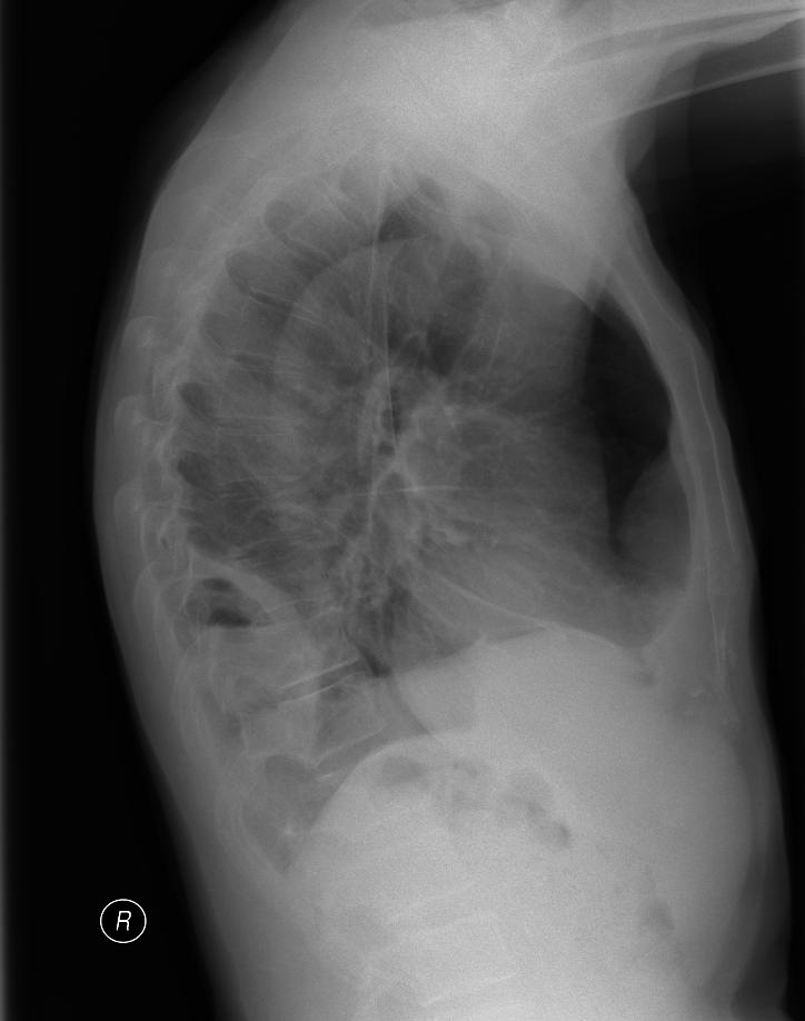
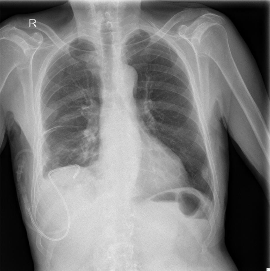
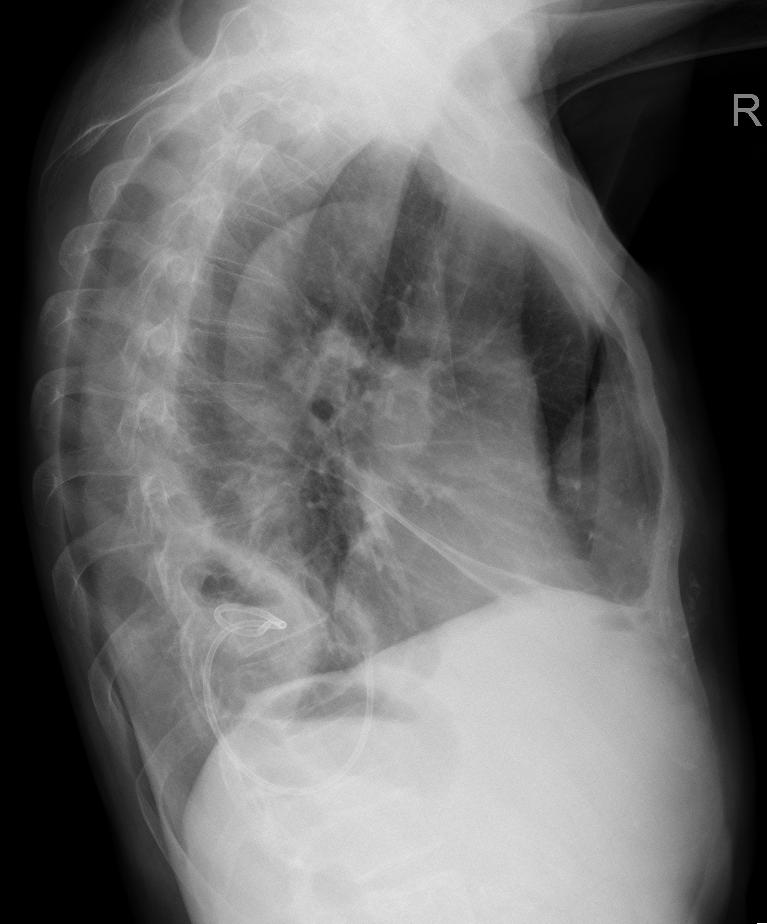
The patient was sent due to cavity lesions with fluid levels on the right being found according to RTG.
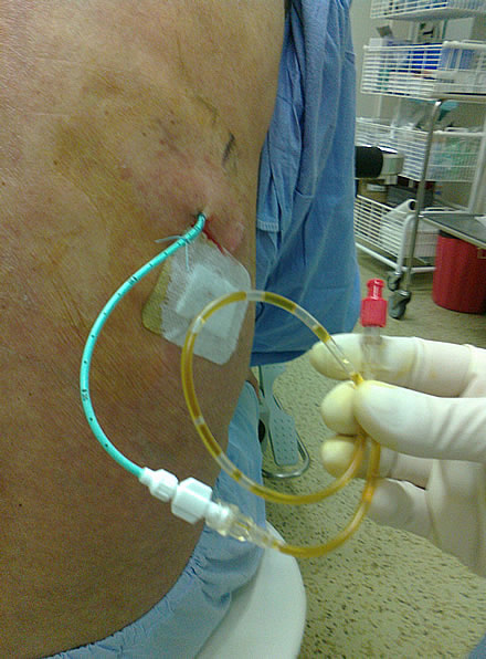
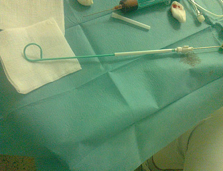
The ultrasound shows an encapsulated collection of fluid on the right. Purulent exudate was collected when the fluid was punctured, so under ultrasound guidance a pig-tail drain (pictured) was inserted
and after taking out the metal mandrin the twisted end of the pig-tail drain can be seen on the
ultrasound. After flushing it out, the encapsulated collection was resorbed. Cytology of the effusion confirmed empyema.
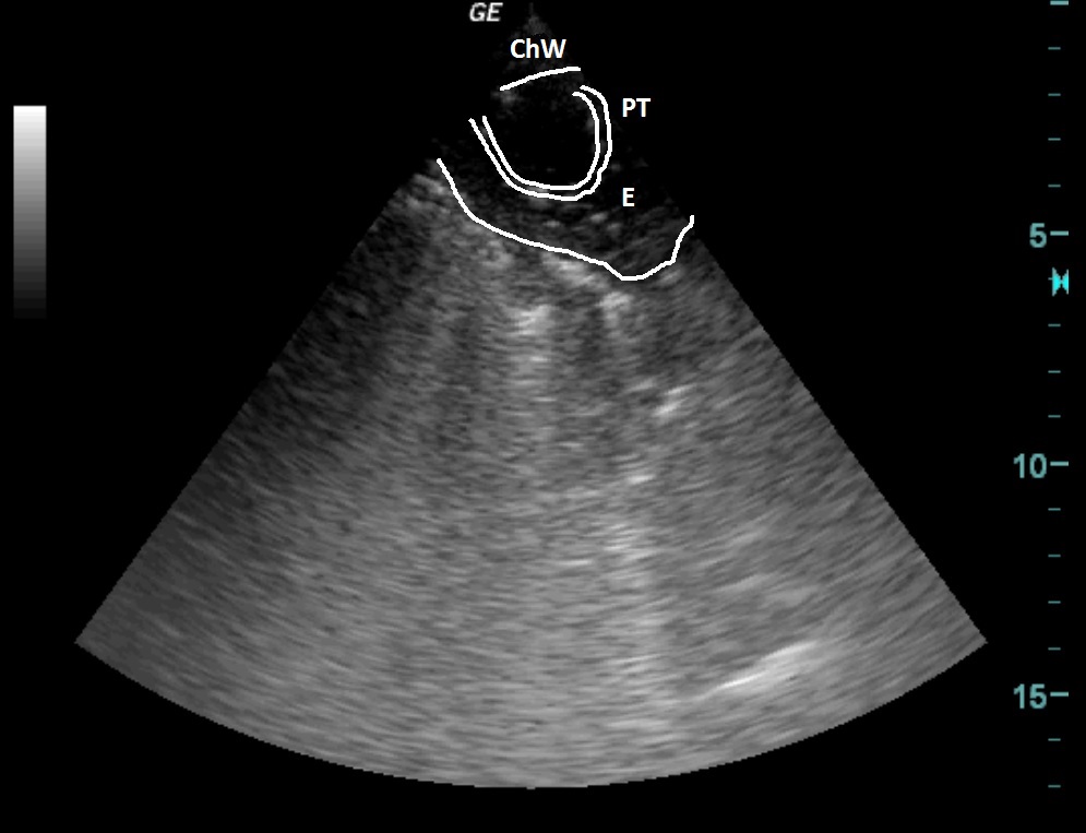
| ChW | - | Chest Wall |
| PT | - | Pigtail drain |
| E | - | Empyema |
After drainage

 česky
česky english