Sono Atlas

   |   

Your browser does not support images in SVG format, please update your browser.

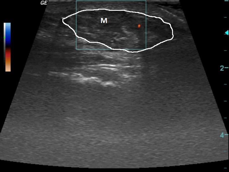
A patient with verified small cell neuroendocrine lung cancer, metastasis in the adrenal gland, the brain and the subcutaneous tissue of the chest wall on the right.

M     -   metastasis

On the ultrasound we can see an oval inhomogeneous structure in the subcutaneous tissue of the chest wall, and with colour Doppler we can see vascularization.


RTG
 

  © MUDr. Rastislav Šimek, 2013

  Project Partner:
Logo GlaxoSmithKline

X-ray and CT documentation used - Department of Imaging Methods, Tomas Bata Regional Hospital, JSC, Zlín.
All materials used on this site are for educational purposes and their use, modification, distribution or use in any other way either partially or completely without the prior written consent of the author is prohibited.