|
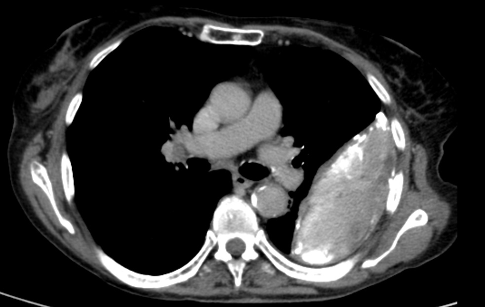
A patient with soft-tissue collection in the left hemithorax found by CT. Hospitalised for further treatment.
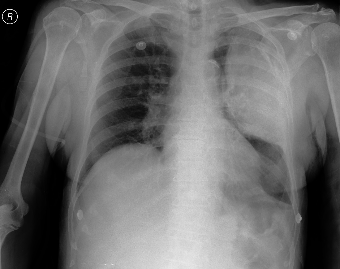
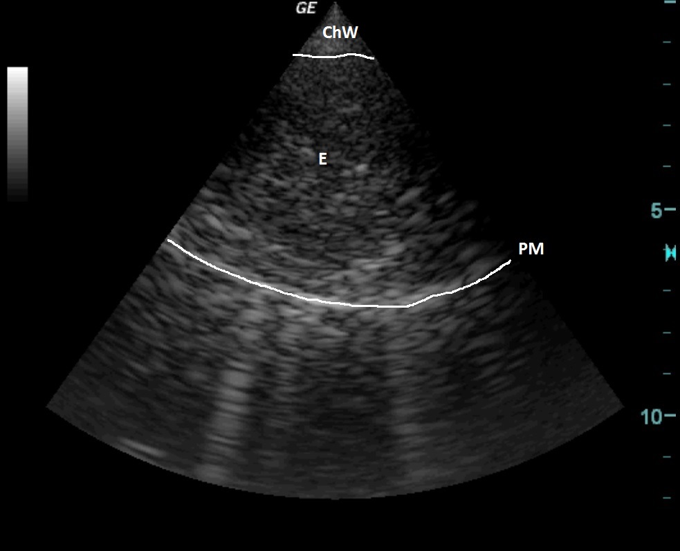
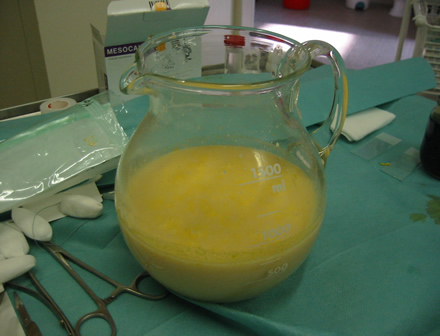
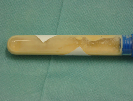
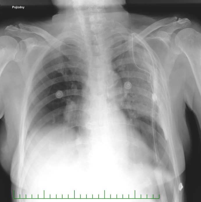
The ultrasound examination showed an encapsulated mass with hyperechoic structures. Pleural biopsy performed, which confirmed thick purulent exudate; cytology confirmed empyema. This was followed by thoracic drainage under ultrasound guidance (see X-ray after drainage), drain inserted on the dorsal side through a parascapular flap. The dense sedimenting fluid was removed (see picture).
| ChW | - | Chest Wall |
| PM | - | Pyogenic Membrane |
| E | - | Empyema |
 česky
česky english