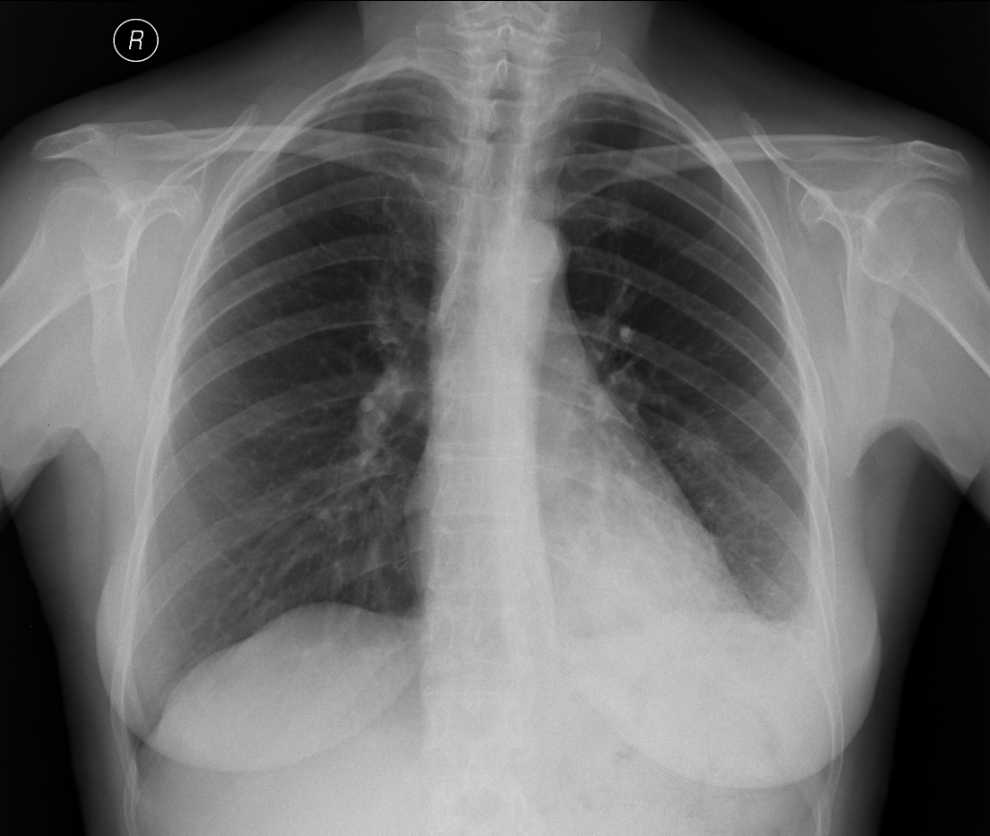
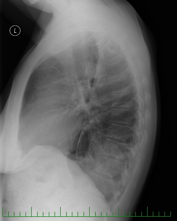
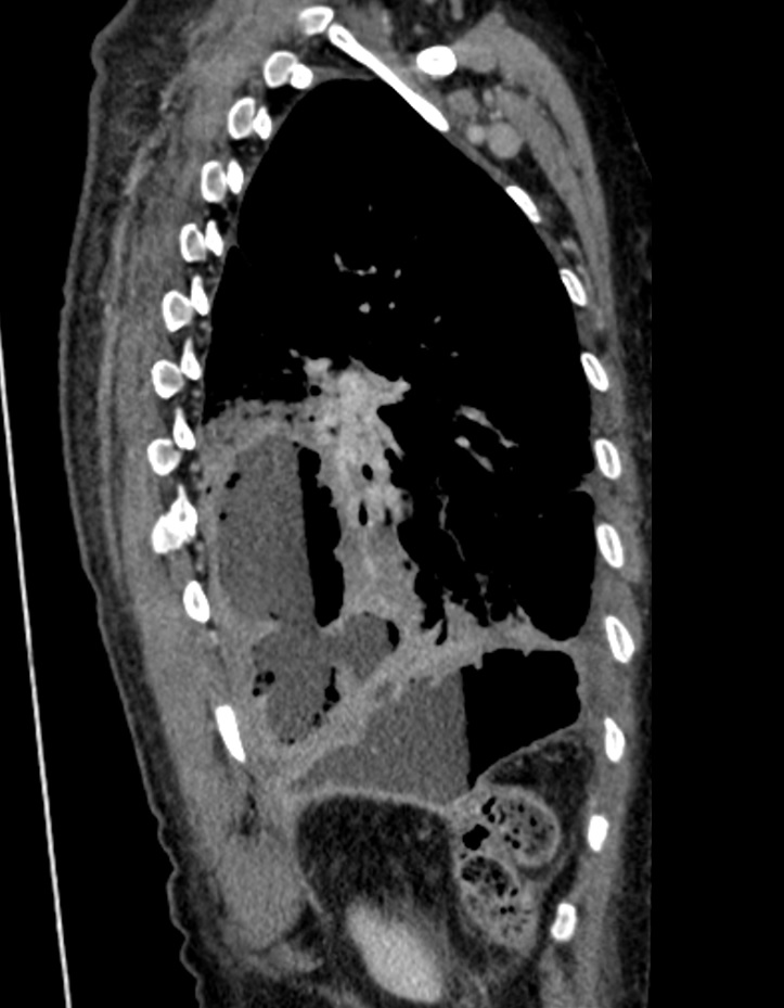
The patient has a malignant retroperitoneal tumour and has already had surgery. She was sent for further investigation due to an ambiguous finding from the X-ray of the left side of the chest, where there is an indistinct shaded area in the sideways projection of the rear diaphragmatic angle.
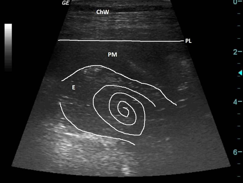
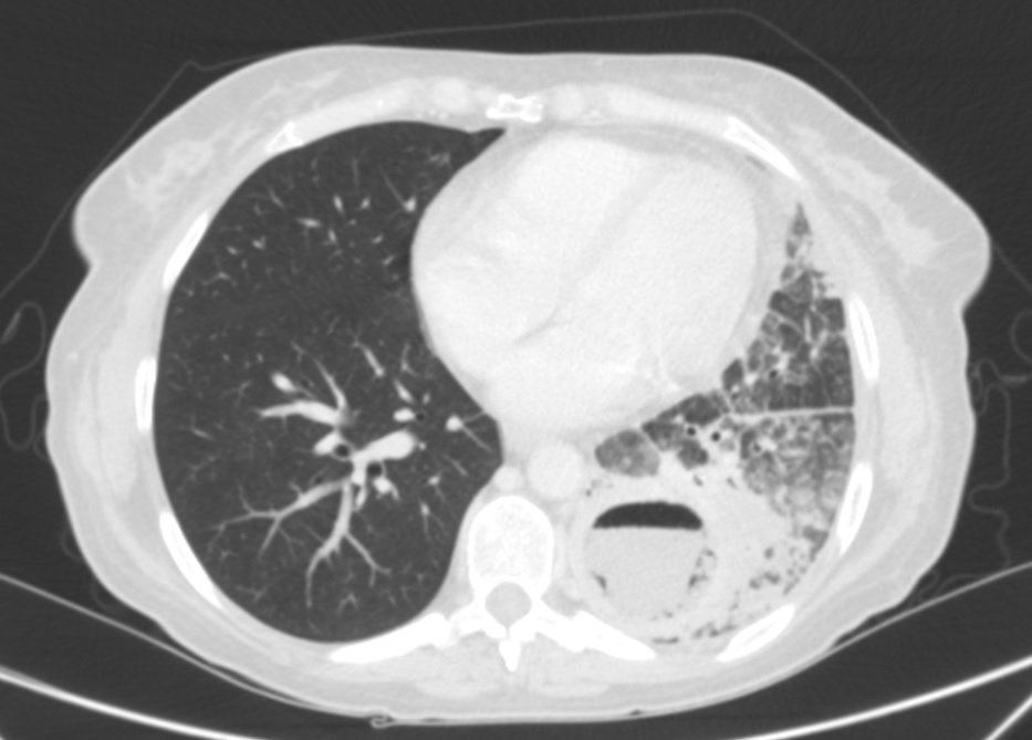
An ultrasonographic examination of the chest was performed, where a hyperechoic fluid collection, which swirls when breathing, is encapsulated, inflammatory collection suspected. It is thus complemented by CT, where we see a cavity lesion with fluid levels of free fluid with the character of an abscess, inflammatory changes in the lung interstitium, delimited collection under the left diaphragm, diaphragm defect and strong suspicion of communication between the two collections. Left subphrenic drainage collection performed surgically, bad-smelling macroscopically cloudy fluid, cytology cellular debris. After antibiotic therapy almost complete regression of the findings.
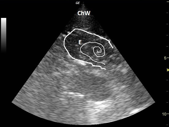
| ChW | - | Chest Wall |
| E | - | Empyema |
| PM | - | Pyogenic membrane |
| PL | - | Pleural line |
 česky
česky english