|
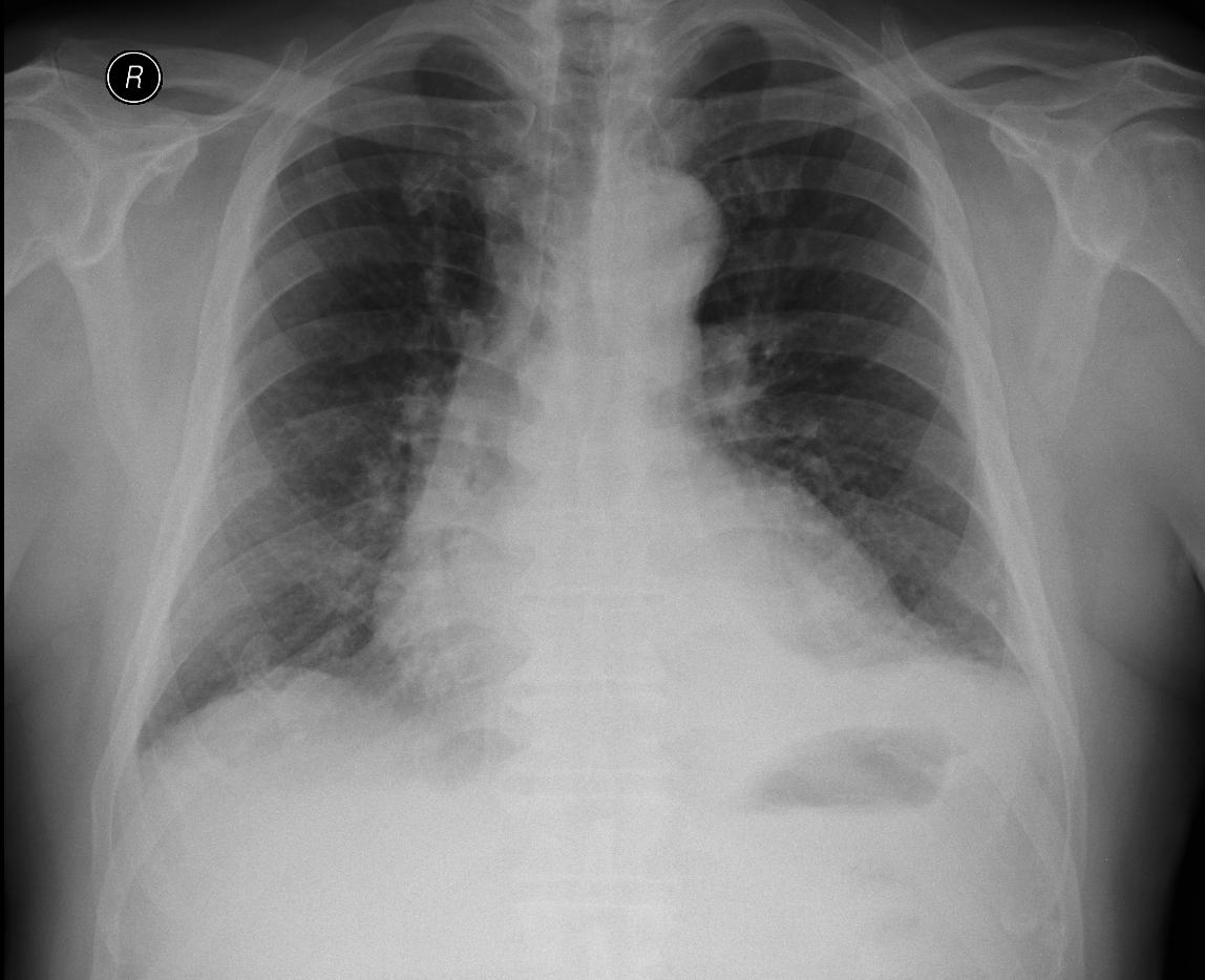
A patient with bilateral effusion, sent for verification.
Under ultrasonographic guidance a diagnostic pleural puncture on the right was performed, and 20 ml of straw-yellow exudate was extracted, biochemical character of exudate, cytology mesothelial type effusion. On the video we see small fluidothorax and the puncture needle, movement of the compressed lung parenchyma when breathing. If necessary, a sonographic check-up of the puncture needle can be done and it is possible to change the needle position as required.
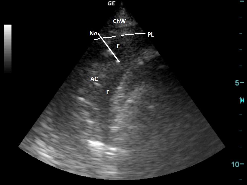
| ChW | - | Chest Wall |
| F | - | Fluidothoraxx |
| Ne | - | Puncture Needle |
| PL | - | Pleural Line |
| AC | - | Compressive atelectasis of the lung parenchyma |
 česky
česky english